
COVID-19, Lung Ultrasound
September 20, 2020
Intraoperative Vascular Ultrasound
September 22, 2020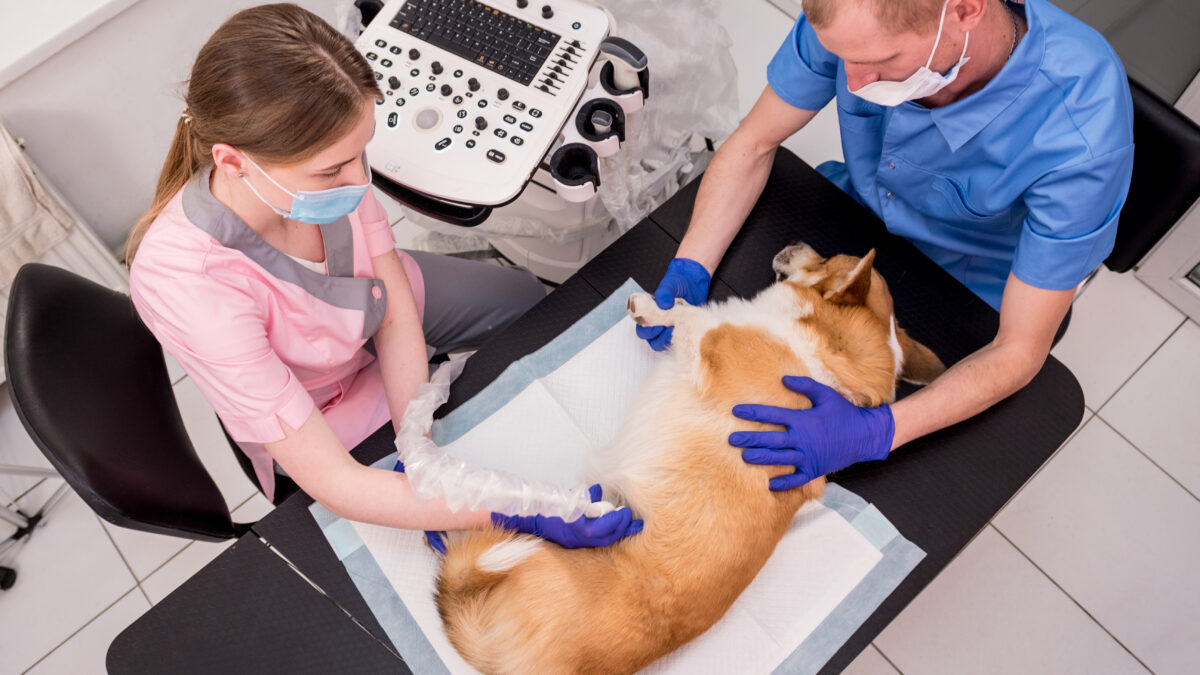
Pet ultrasounds are a safe and commonly used way of imaging the inside of dogs, cats, and other pets. They’re a non-invasive method of imaging, which uses sound waves to help vets visualize a pet’s internal organs and diagnose conditions such as those affecting the heart, liver, kidneys, and bladder.
Ultrasound gives us data as to whether the structure of a pet’s organ is as it ought to be and is practically operating; for instance to appear where blood is flowing and how the heart is contracting. It is a very secure technology for pets, as sound waves do not harm or affect the body during the procedure.
Which Ultrasound Scanner is the best for Pets?
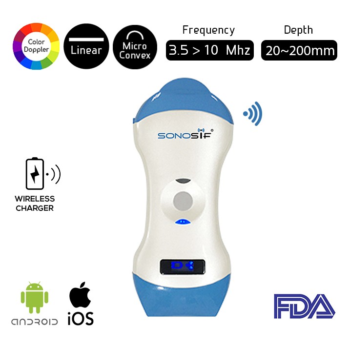
Ultrasound can identify an issue profound inside tissue or an organ without obtrusive surgery. On the off chance that a test of tissue is required, an ultrasound-guided biopsy can be performed after the pet has been given suitable anesthetics. Accordingly, SONOSIF’s Research and Development team recommends the wireless Double head Ultrasound scanner Micro convex and linear MCL1-CD to vets and radiologists.
A micro-convex probe is valuable within the scanning of little animals. Usually for two fundamental reasons; to begin with, the animal’s abdomen is little which makes a wide probe less practical. Second, the ultrasound waves don’t have to be travel distant exceptionally into the body, so high-frequency soundwaves would be appropriate to deliver way better quality sound waves.
Depending on the images delivered, ultrasound can take different shapes. In veterinary work, B-mode(brightness-mode) ultrasound, a more commonly called 2-dimensional ultrasound is the foremost common shape. This gives a two-dimensional picture of the organ checked. Typically the sort of ultrasound that’s utilized to examine abdominal structures, perform pregnancy diagnosis, assess cardiac function, and look at the eyes for certain eye infections.
A bit like in Humans, a pets ultrasound scanner can be utilized to see at the uterus. For instance, at the puppies and kittens living there. This permits the specialist to see at which arrange is the pregnancy, and evaluate how healthy the babies are. Indeed more vitally, it makes a difference look at the uterus in a debilitated animal to decide whether or not she has a Pyo( possibly deadly womb infection). Here, by seeking out for two dark circles ( in some cases called ” shotgun barrels”) which are the horns of the uterus when they are filled with liquid.
It is additionally utilized to look at other abdominal organs such as the kidneys, the bladder, the spleen, and the liver. This way, tumors, twists, and other wounds can be recognized without having to open up the patient in surgery.
The scanner can be used also to see on the off chance that there’s free blood or fluid inside the abdomen that might show inner bleeding or guide a biopsy needle to a suspicious knot, without requiring surgery.
Furthermore, We can use the Ultrasound scanner at the cardiac level which usually referred to as echocardiography. Doppler ultrasound may be a specialized shape of cardiac ultrasound in which the direction and speed of the bloodstream within the heart and blood vessels can be measures. Color-flow doppler technology makes it indeed less demanding to watch the flow of blood through the heart and vital blood vessels. Moreover, it allows a look at the beating heart of the cat, dog, or any pet.
Most importantly, an Ultrasound scanner can diagnose a wide range of medical conditions such as abnormalities of the gall bladder, urinary bladder, prostate or kidneys, abnormal blood vessels, uterine infections, fluid around the heart (pericardial effusion), etc…

