- Immediate contact :
- +1-323-988-5889
- info@sonosif.com

Laparoscopic Ultrasonography
September 22, 2020
Ultrasound Scanner for Bladder Diverticulum
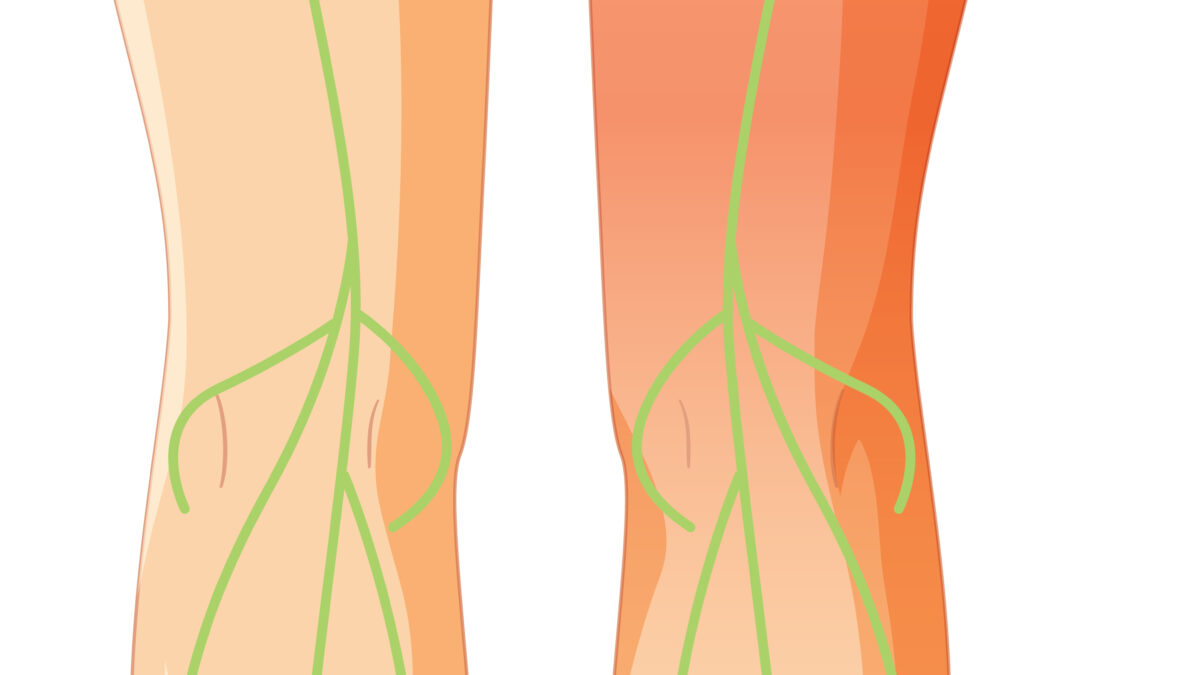
September 24, 2020The lateral femoral cutaneous nerve (LFCN) is a sensory nerve that provides sensation to the outer and frontal side of the thigh just above the greater trochanter to the knee. It divides into several branches innervating the lateral and anterior aspects of the thigh. LFCN‘s variable anatomy makes it challenging to perform an effective landmark-based block.
LFCN has received much attention because of its association with Meralgia paresthetica, a condition characterized by tingling, numbness, and burning pain in the outer thigh.
Knowledge of the lateral femoral cutaneous nerve’s anatomical variations are crucial for preventing nerve injury during the insertion of needles into the anterior superior iliac spine (ASIS) and other surgical procedures.
A lateral femoral cutaneous nerve block is an injection of a local anesthetic and steroid to block the nerves that influence pain in the upper leg. It is useful in the evaluation and management of lateral thigh pain.
According to researches, the rate of successful anesthesia based on the use of anatomical landmarks has only been approximately 40%. An Ultrasound-Guided Nerve Block Technique, however, was proven to have more accurate needle insertion into the appropriate fascial plane through which the LFCN passes
Which Ultrasound Scanner is best for Lateral Femoral Cutaneous Nerve?
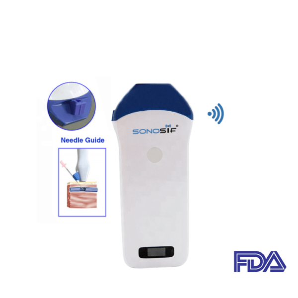
Radiologists, anesthesiologists, neurologists, and surgeons will need the Mini Linear Handheld WiFi Ultrasound Scanner MLCD
There are several promising studies about the ability of ultrasound in the evaluation of the LFCN, the use of ultrasound guidance in regional anesthesia for blocking the LFCN and the use of ultrasound in nerve conduction studies of the LFCN.
The advantage of Ultrasound-guided procedures is the control of the needle insertion path. The practitioner can visualize the needle in real-time as it enters the body and traverses to the desired location. This assures that the medication is accurately injected at the intended site.
MLCD offers High-resolution ultrasound images and High intensity focused digital thanks to its higher frequency (10MHz / 12MHz / 14MHz.). This allows the practitioner to identify the LFCN if the intermuscular space between the tensor fasciae latae muscle and the sartorius is used as an initial sonographic landmark.
The device comes with a needle guide holder. Hence, it can be directly set to the guide pin frame. Coupled with the software that can quickly locate the depth and diameter of puncture’s navigation
The Mini Linear Handheld WiFi Ultrasound Scanner MLCD also enables clinicians and patients to see the impact of treatments often before visual signs are apparent.
In addition, MLCD is a wireless device and it is iOS and Android compatible to give superior image quality. Thanks to its lightweight (250g) and small design it is easy to carry and use.
Disclaimer: Although the information we provide is used by different doctors and medical staff to perform their procedures and clinical applications, the information contained in this article is for consideration only. SONOSIF is not responsible neither for the misuse of the device nor for the wrong or random generalizability of the device in all clinical applications or procedures mentioned in our articles. Users must have the proper training and skills to perform the procedure with each ultrasound scanner device.
The products mentioned in this article are only for sale to medical staff (doctors, nurses, certified practitioners, etc.) or to private users assisted by or under the supervision of a medical professional.


