
Transrectal Prostate Ultrasound (TRUS)
October 20, 2020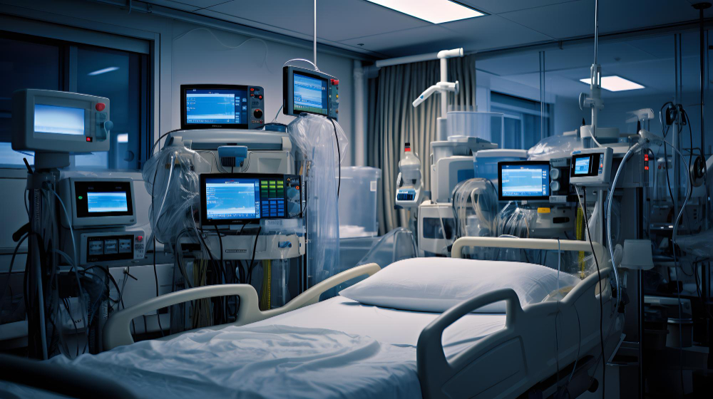
Focused Cardiac Ultrasound: FOCUS
October 21, 2020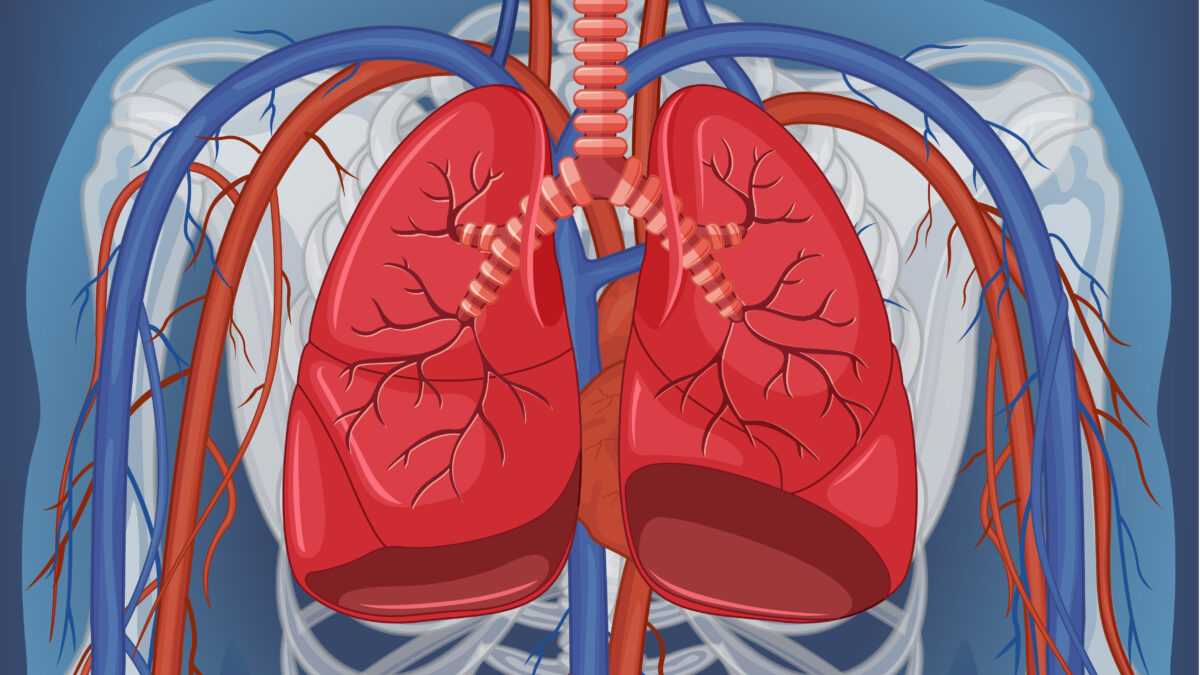
Thoracentesis is a procedure in which a needle is inserted into the pleural space between the lungs and the chest wall to remove excess fluid from the pleural space to help the patient breathe easier. It may be done to determine the cause of the pleural effusion. Some conditions such as heart failure, lung infections, and tumors can cause pleural effusions.
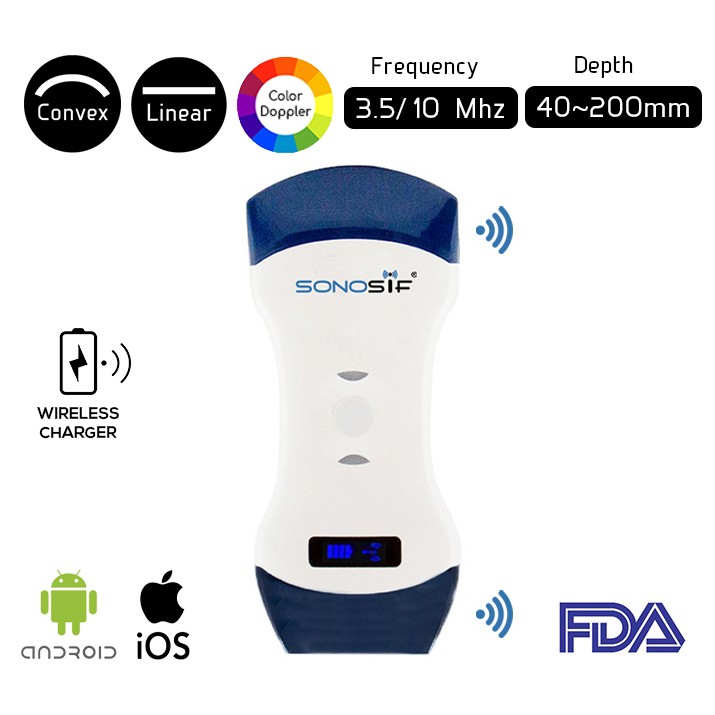
Which Ultrasound Scanner is suitable for the Thoracentesis assessment?
The Convex and Linear Color Doppler Wi-Fi Double Head Ultrasound Scanner CLCD is highly recommended to our Pulmonologists, intensivists, and Emergency medicine doctors clients.
This Device has two heads. Thus, making it more practical and more affordable than buying two separate single-headed probes. The Linear side of the Doppler allows you to evaluate the more superficial parts of the body while the Convex part is used for in-depth examinations.
A curvilinear probe with a frequency of 3.5–5.0 MHz is best suited for performing a thoracentesis. This allows for better visualization of deeper structures and is more than adequate for visualizing more superficial structures adjacent to the chest wall. A higher-frequency probe between 5.0 and 7.0 MHz is effective for visualizing chest wall structures and the parietal pleura.
Real-time ultrasound-guided thoracentesis is a safe procedure with a low complication rate compared to that for blind techniques, has a complication rate similar to that for other ultrasound-guided techniques, and leads to significant improvement in oxygenation rates.
The Ultrasound is done to ascertain the correct area where the needle will go. It guides the practitioner while inserting the needle or tube below the ribs into the pleural space. The excess fluid will then be drained out.
The ultrasound scanner provides improved needle insertion site selection, better characterization of pleural anatomy, and higher rates of procedural success than physical examination alone.
To conclude Ultrasound can precisely identify the location of the fluid so that the chest wall can be marked in preparation for thoracentesis. Moreover, it helps to follow-up the procedure and checks the results.
References: Real-time ultrasound-guided thoracentesis, Thoracentesis,
Disclaimer: Although the information we provide is used by different doctors and medical staff to perform their procedures and clinical applications, the information contained in this article is for consideration only. SONOSIF is not responsible neither for the misuse of the device nor for the wrong or random generalizability of the device in all clinical applications or procedures mentioned in our articles. Users must have the proper training and skills to perform the procedure with each ultrasound scanner device.
The products mentioned in this article are only for sale to medical staff (doctors, nurses, certified practitioners, etc.) or to private users assisted by or under the supervision of a medical professional.


