
Veterinary: Bovine Ultrasound examination
June 2, 2021
Fetal Ultrasound
June 8, 2021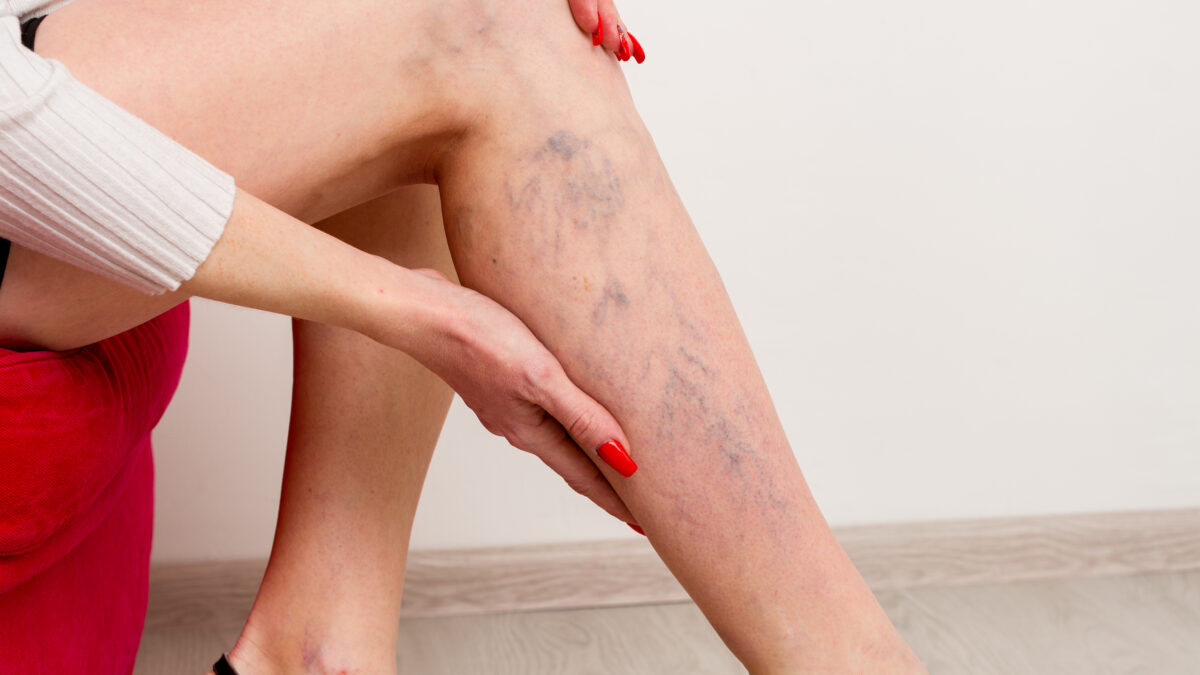
Endovenous laser ablation (EVLA) is a minimally invasive treatment for leg varicosities that is widely used and extremely successful.
Varicose veins are bulging, palpable, and convoluted veins that are visible underneath the skin and have a diameter more than 3-4 mm. varicose veins most typically affect the lower limbs .
A high percentage of the varicose veins on the legs are due to superficial venous deficits of the large saphenous vein, but in the majority of the individuals with superficial venous deficiency, the saphenofemoral junction is the major source of reflux.
Thus, Endovenous Laser Ablation (EVLA) should be performed in order to close off a varicose or an enlarged swollen vein
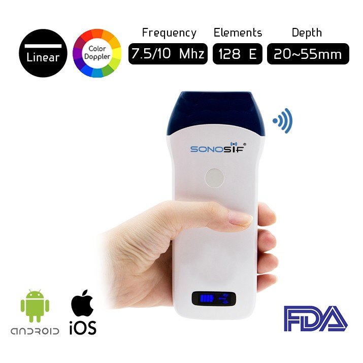
Using ultrasonic scanning is a major non-invasive study used to examine vascular issues such as varicose veins. It is used to assess the characteristics and condition of the veins of the patient.
For instance, the Wireless Color Doppler Linear Ultrasound Scanner L2CD is highly recommended since it has a high frequency of 7.5 to 10 MHz which aids in detecting the veins and the points where they intersect.
With L2CD real-time guidance , a laser fibre is placed into the abnormal vein through a tiny incision. The vein is then numbed with local anaesthetic, and the laser activated as the fibre is slowly removed. This produces a reaction in the vein wall along the treated section, resulting in collapse and sclerosis of the vein wall with minimal discomfort.
To sum up, Ultrasound-guided EVLA of lower limb varicose veins is an effective way that leads to a satisfying clinical outcome. It is a minimally invasive and safe interventional procedure, related to a low risk of complications.
References: Endovenous laser ablation (EVLA): a review of mechanisms, modeling outcomes, and issues for debate, Ultrasound Guided (US) Endovenous Laser Ablation (EVLA) of Lower Limb Varicose Veins: How to Do
Disclaimer: Although the information we provide is used by different doctors and medical staff to perform their procedures and clinical applications, the information contained in this article is for consideration only. SONOSIF is not responsible neither for the misuse of the device nor for the wrong or random generalizability of the device in all clinical applications or procedures mentioned in our articles. Users must have the proper training and skills to perform the procedure with each ultrasound scanner device.
The products mentioned in this article are only for sale to medical staff (doctors, nurses, certified practitioners, etc.) or to private users assisted by or under the supervision of a medical professional.


