
Abnormal Appendix: Appendicitis
October 20, 2020
Transrectal Prostate Ultrasound (TRUS)
October 20, 2020
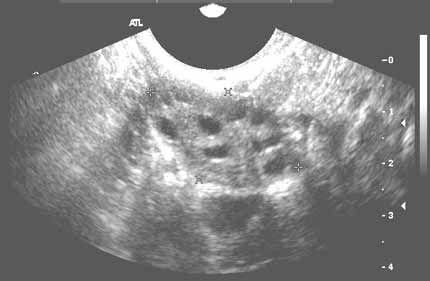
Abdominal Pelvic Scan a noninvasive diagnostic exam that produces images that are used to assess organs and structures within the female pelvis. It allows quick visualization of the female pelvic organs and structures including the uterus, cervix, vagina, fallopian tubes, and ovaries.
Which ultrasound Scanner is best used for Abdominal Pelvic Scan?
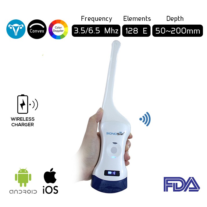
Since Pelvic imaging may be performed using either Transabdominal trasnducer or Transvaginal transducer; or using both methods, The Convex and Transvaginal Color Doppler Double Head Wi-Fi Ultrasound Scanner CTC-3.1 is highly recommended to our gynechologist and obstetrician clients. In which, It has two heads. Thus, making it more practical and more affordable than buying two separate single headed probes.
Pelvic Ultrasound is used for measurement and evaluation of female pelvic organs. Yet, the assessment may include Size, shape, and position of the uterus and ovaries, Thickness, echogenicity (darkness or lightness of the image related to the density of the tissue), and presence of fluids or masses in the endometrium, myometrium (uterine muscle tissue), fallopian tubes, or in or near the bladder, Length and thickness of the cervix, Changes in bladder shape, and Blood flow through pelvic organs.
In other words, Pelvic ultrasound can provide much information about the size, location, and structure of pelvic masses.
Another type of ultrasound used for pelvic examination is Doppler ultrasound, sometimes called a duplex study, used to show the speed and direction of blood flow in certain pelvic organs. Unlike a standard ultrasound, some sound waves during the Doppler exam are audible.
Ultrasound may also be used to assist with other procedures such as endometrial biopsy . Transvaginal ultrasound may be used with sonohysterography, a procedure in which the uterus is filled with fluid to distend it for better imaging.
To sum up, Abdominal Pelvic Scan provides qualitative and quantitative data about the female pelvic.
References: Pelvic Ulrasound.
Disclaimer: Although the information we provide is used by different doctors and medical staff to perform their procedures and clinical applications, the information contained in this article is for consideration only. SONOSIF is not responsible neither for the misuse of the device nor for the wrong or random generalizability of the device in all clinical applications or procedures mentioned in our articles. Users must have the proper training and skills to perform the procedure with each ultrasound scanner device.
The products mentioned in this article are only for sale to medical staff (doctors, nurses, certified practitioners, etc.) or to private users assisted by or under the supervision of a medical professional.


