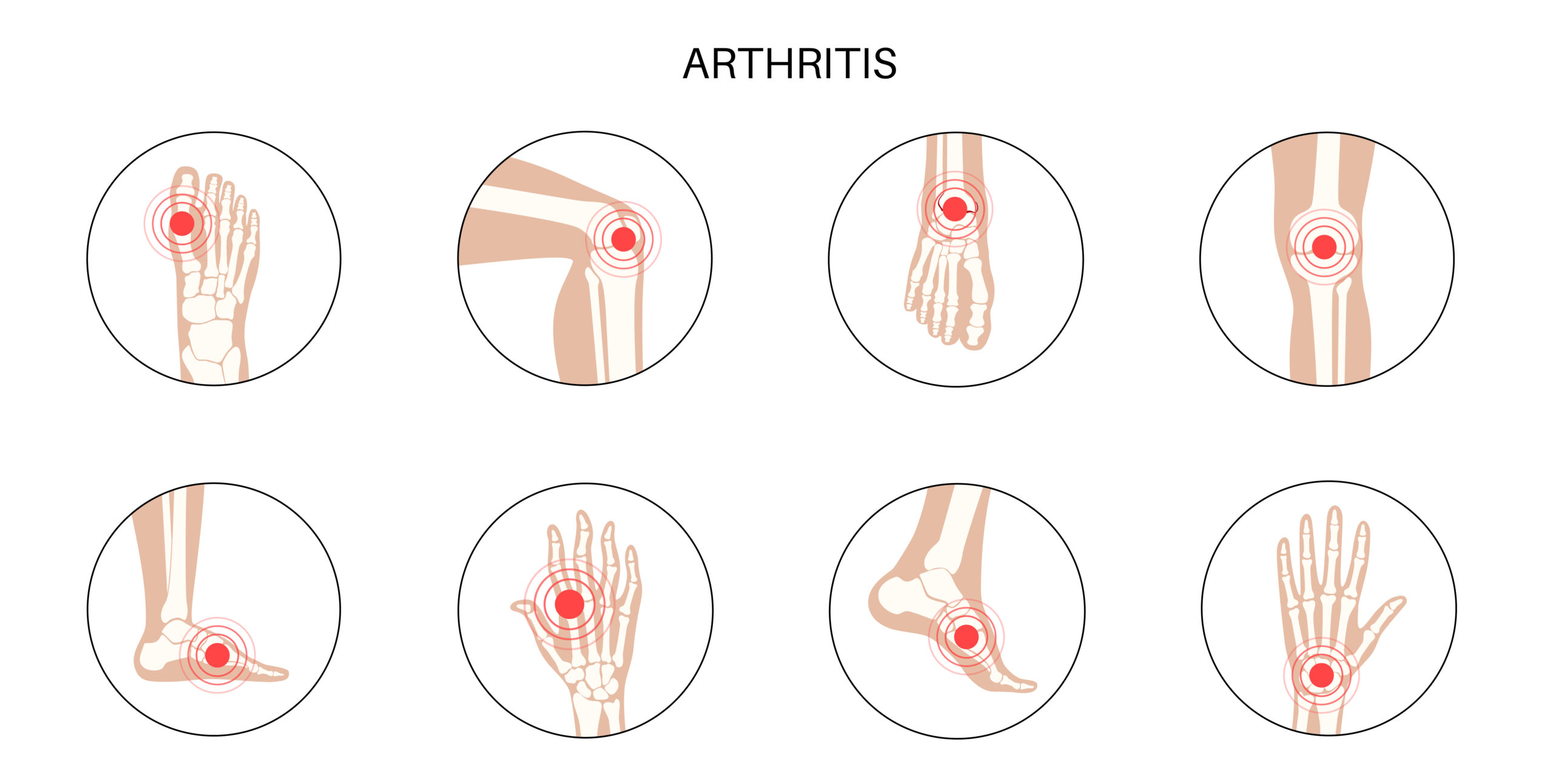
Ultrasound-guided Arthrocentesis
October 9, 2020
Identifying Perforators in Reconstructive Surgery
October 11, 2020Airway Assessment is a procedure done to identify potential problems with the maintenance of oxygenation and ventilation during airway management. It is the first step in formulating an appropriate airway plan.
Ultrasound imaging technique has emerged as a simple, portable, and non-invasive tool helpful for airway assessment and management. It can visualize anatomical structures in the supraglottic, glottic, and subglottic regions.
Which Ultrasound Scanner is best for Airway Assessment?
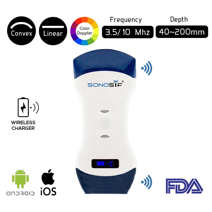
The Convex and Linear Color Doppler Wi-Fi Double Head Ultrasound Scanner CLCD is strongly suggested to anesthesiologist clients. It is a reliable tool for the assessment of the subglottic airway. The CLCD allows the doctor to estimate the appropriate tracheostomy the size and length and avoid anterior neck structures as well as posterior tracheal wall injuries.
The curved and Linear probes are commonly used for this assessment. A high-frequency linear probe 7.5 MHz and 5 MHz curved array probe are used for visualization of superficial and deeper structures of the airway.
Reflection, refraction, scatter, absorption, and transmission of sound occur as it passes through soft tissue structures, allowing characterization of the shape and internal architecture of that structure in addition to those behind it. Reflection of sound is marked at interfaces between tissues of different acoustic impedance. The image is built from the reflected sound signals.
Reflection, refraction, scatter, absorption, and transmission of sound occur as it passes through soft tissue structures, allowing characterization of the shape and internal architecture of that structure in addition to those behind it. Reflection of sound is marked at interfaces between tissues of different acoustic impedance. The image is built from the reflected sound signals.
As for the curved low-frequency probe, It is used to visualize the tongue and shadows of the hyoid bone and mandible with the patient in the supine position. The hyomental distances are measured from the upper border of the hyoid bone to the lower border of the mentum in the neutral and extended head positions, respectively.
To sum up, Because of high-quality imaging, non-invasiveness, and relatively low cost, ultrasonography has been utilized as a valuable adjunct to the clinical assessment of the airway.
References: Airway Assessment, Point-of-care ultrasound in the airway assessment, Ultrasound of the airway


