Pets Ultrasound Scanning
September 22, 2020
Laparoscopic Ultrasonography
September 22, 2020An assortment of intraoperative imaging and evaluation strategies are accessible in modern operating rooms. However, there is no standardized strategy for surveying and assessing vascular procedures.
The doctor may instruct an ultrasound to view and record the blood flow immediately during a surgical bypass procedure (repairing diseased arteries in the neck, kidneys, or lower legs with healthy veins from the leg).
Which ultrasound is used for Intraoperative Vascular Ultrasound?
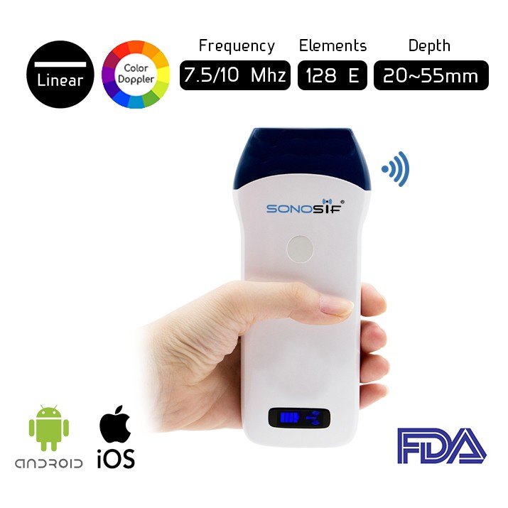
The use of high-quality images offers critical knowledge and increases surgeon confidence, playing an important role in any surgery and may help reduce the rate of reoperation in some cases. For this reason, SONOSIF’s Research and Development team always recommends the wireless color Doppler Linear Ultrasound Scanner L2CD to our physicians and vascular surgeons clients.
Yet, a direct 7.5MHz is utilized in vascular diagnosis intraoperative. The vascular specialist performs the ultrasound method after surgery but before the skin is closed. The L2CD is set in a sterile on the assigned area.
During the surgery, the practitioner holds the transducer following the blood vessel being assessed. Sound waves bounce off the blood moving within the arteries and the tissue within the body This makes echoes that reflect the transducer.
The use of Intraoperative sonography can offer assistance to record the technical success of the procedure and to distinguish possibly correctable thromboembolic sources that may lead to perioperative stroke or restenosis.
In addition, routine intraoperative duplex ultrasound examination of the carotid reconstruction permits early diagnosis and prompt adjustment of morphologic as well as hemodynamic injuries.
Last but not least, as the contrast in transit times of the ultrasound beams is based only on moving components within the blood vessels, the estimations are not affected by the inward distance across the vessel wall. This is often significant for atherosclerotic arteries.
References: Intraoperative Duplex Ultrasound, Intraoperative use of Vascular Ultrasound.


