
LUS: Lung Ultrasound
September 30, 2020
Musculoskeletal Ultrasound
October 1, 2020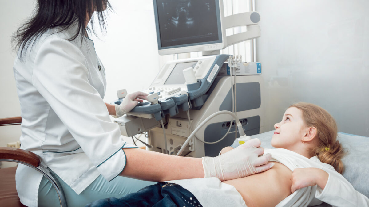
Pediatric Ultrasound uses sound waves to produce images inside the body. It does not use radiation and has no side effects. It is very useful in assessing the causes of abdominal, pelvic, or excruciating pain in children.
Ultrasound can not only detect the cause of abdominal pain in pediatric patients, but it can also help doctors to formulate appropriate treatment plans. Ultrasound examination offers the advantages of fast, simple, safe, and accurate diagnosis. This is the first option for diagnosing abdominal pain in children.
Ultrasound is the most important imaging technique in pediatrics; more than any other branch of medicine. This is due to several factors. X-ray exposure is a more important factor for pediatric patients than adults with an X-ray dose, and thus, pediatric patients are more likely to receive radiation. In addition, certain types of immature tissue are vulnerable to radiation. Thus it is necessary to avoid X-ray use in children and young adults.
Which Ultrasound is the best for Pediatric diagnosis?
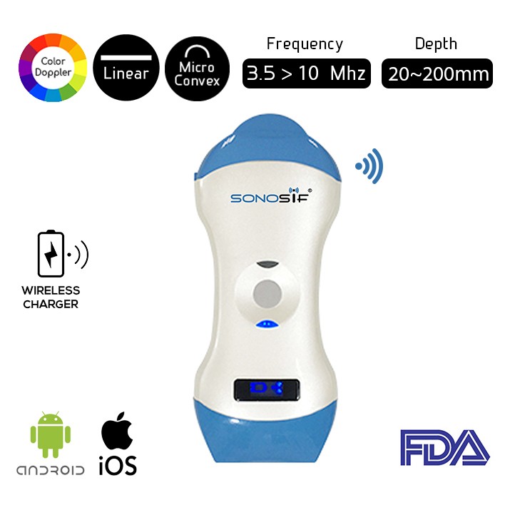
Using a high-frequency ultrasound Wireless Double head Ultrasound Scanner Micro-convex and Linear MCL1-CD is highly recommended by our SONOSIF ‘s Research and Development team to our pediatric specialists and radiologists clients.
In fact, high-frequency ultrasound scanner MCL1-CD double head (ideal for relatively shallow penetration depth-based neonates) often produces image quality that is unmatched in any other. This type of ultrasound scanner offers high resolution. Yet, in some cases, it can only detect congenital heart defects, brain morphology, cranial bleeding, and hemodynamic complications.
Numerous pediatric viral diseases that cause the formation of debris or thrombus in the superficial and deep veins can be detected by high-resolution ultrasound. Other applications include improved neurotransmitter properties, intima-media thickness of large and small vessels, internal contents of peripheral veins, and salivary glands. Thyroid nodules, superficial vascular disorders, and ocular pathology, especially in young children, will also be best assessed through high-resolution imaging.
Ultrasound is widely available and can be used for screening purposes. For example, the current practice of hip screening for all newborns has reduced the number of hip defects required for surgery.
To sum up, it is important to note that to check for superficial soft tissue abnormalities in young children, ultrasound provides a higher local resolution than MRI. The high-resolution ultrasound scanner’s local resolution is in micrometers, while MRI is in millimeters. Within the availability of FDA-approved ultrasound contrast, it can now provide information on the dynamic growth of enhanced MRI in contrast to multiple caps.
References: Children’s (pediatric) Ultrasound, The advantages of Pediatric ultrasound,


