- Immediate contact :
- +1-323-988-5889
- info@sonosif.com

GUS: Gastric Ultrasound Scanner
October 27, 2020
Ultrasound Scanner for Polycystic Ovarian Syndrome Assessment
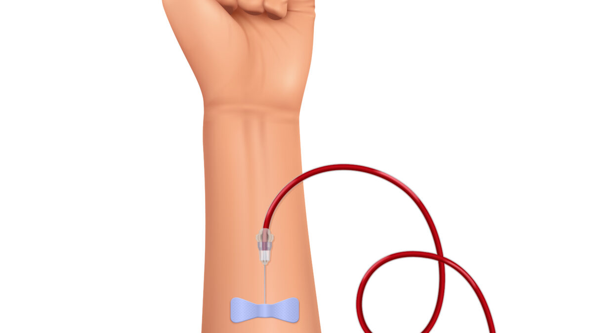
January 12, 2021Peripherally Inserted Central Catheter (PICC) is a thin flexible tube that is inserted into a vein upper arm and guided into a large vein above the right side of the heart called the superior vena cava. It is used to give intravenous fluids, blood transfusions, chemotherapy, etc…
PICCs are inserted by ultrasound-guided puncture and cannulation of the deep upper arm veins (most often the basilic or brachial vein) via the modified Seldinger technique (venipuncture with a small-gauge needle, insertion of a thin guidewire through the needle, removal of the needle, insertion of a micro-introducer-dilator over the guidewire, removal of the wire and dilator, insertion of the catheter through the introducer).
This technique facilitates the catheterization of large-bore veins of known diameter and trajectory while allowing positioning of the exit site in the upper part of the arm (halfway between the elbow and the axilla).
As a final result, the rate of insertion failure is minimal, insertion-related complications are almost negligible, the exit site is handled easily by patients and nursing staff, and the rate of late complications (infection, catheter-related venous thrombosis, dislocation) is low.
Which Ultrasound Scanner is suitable for PICC?
The High-frequency Mini-Linear Handheld Wi-Fi Ultrasound Scanner MLCD is highly recommended to Anesthesiologists, Radiologists, Radiology physician assistants, and Nurses.
Ultrasound-guided puncture is most commonly performed via an “out-of-plane” approach (the needle trajectory does not lie in the transducer plane).
When the transducers are equipped with a needle holder, it facilitates puncture.
Moreover, When using ultrasound for venipuncture, position the ultrasound handheld probe (also called a transducer) on the skin to produce either transverse or sagittal images of veins.
When you position the probe perpendicular to a vein, the vein appears as a circle on the ultrasound monitor screen; this is a transverse view.
Placing the probe parallel to the vein produces a sagittal (longitudinal) view of the vein.
References: Ultrasound-guided placement of peripherally inserted central venous catheters, PICC,
Disclaimer: Although the information we provide is used by different doctors and medical staff to perform their procedures and clinical applications, the information contained in this article is for consideration only. SONOSIF is not responsible neither for the misuse of the device nor for the wrong or random generalizability of the device in all clinical applications or procedures mentioned in our articles. Users must have the proper training and skills to perform the procedure with each ultrasound scanner device.
The products mentioned in this article are only for sale to medical staff (doctors, nurses, certified practitioners, etc.) or to private users assisted by or under the supervision of a medical professional.


