Traumatic Extremity Injuries
October 7, 2020
Ultrasound-Guided Thoracic Paravertebral Block
October 8, 2020Pleural drainage is an essential aspect of the management of the critically ill patient in the intensive care unit or accident and emergency department. The primary aim of pleural drainage is the effective drainage of air, blood, or fluids from the pleural space to restore cardiorespiratory function by reexpansion of the lung and the elimination of the mediastinal shift which may cause hemodynamic instability.
Ultrasound guidance during pleural drainage, allows the operator to increase the rate of success of the procedure and reduce its associated risks.
Furthermore, thoracic ultrasound helps clinicians to identify the best puncture site and to guide the drainage insertion procedure. Thoracic ultrasound is essential during these invasive maneuvers to increase safety and decrease potential life-threatening complications. It helps clinicians not only to visualize pleural effusion but also to distinguish between the different types.
Which Ultrasound is suitable for Pleural drainage?
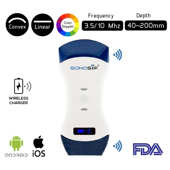
Using a doppler ultrasound is best to visualize intercostal vessels, it helps to prevent vessel injury and ensure a procedure with a low risk of bleeding. Based upon, the Convex and Linear Color Doppler Wi-Fi Double Head Ultrasound Scanner CLCD is highly recommended to our cardiologist, pulmonologist, and thoracic surgeon clients.
A low-frequency transducer from 3.5 to 5 MHz convex is used in the transverse position between two ribs to identify the best puncture site evaluation. Then, the operator can Shift to the high-frequency ultrasound transducer from 7 to 15 MHz. The probe should be used in the transverse position between two ribs to identify the upper and lower borders of the needle insertion area.
The position of the drain is determined by the location and the nature of the collection to be drained, with particular attention to the depth required to reach the pleural fluid and avoid lung injuries. The 5th intercostal space in the mid-axillary line is generally used for most situations. This area is commonly known as the “safe triangle”, bordered by the anterior border of latissimus dorsi, the lateral border of the pectoralis major, a line superior to the horizontal level of the nipple, and an apex below the axilla. The drain should be inserted just above the rib.
The use of bedside ultrasound not only leads to an improvement in the diagnosis but also allows the detection of the best puncture site and the fluid quantification of PLEFF. The positioning of percutaneous pleural drainage with ultrasound guidance increases the procedure’s success rate and safety.
To sum up, Ultrasound’s ease of use, portability to the patient’s bedside, and its accurate examination of the pleural space has allowed for safer pleural procedures such as thoracentesis, chest tube placement, tunneled pleural catheter placement, and medical thoracoscopy.
References: Thoracic Ultrasound for Pleural Effusion in ICU, Chest Tube Drainage
Disclaimer: Although the information we provide is used by different doctors and medical staff to perform their procedures and clinical applications, the information contained in this article is for consideration only. SONOSIF is not responsible neither for the misuse of the device nor for the wrong or random generalizability of the device in all clinical applications or procedures mentioned in our articles. Users must have the proper training and skills to perform the procedure with each ultrasound scanner device.
The products mentioned in this article are only for sale to medical staff (doctors, nurses, certified practitioners, etc.) or to private users assisted by or under the supervision of a medical professional.


