- Immediate contact :
- +1-323-988-5889
- info@sonosif.com

Ultrasound-guided evaluation of the Median nerve at the level of the carpal canal and ulnar nerve
May 22, 2021
Ultrasound Guidance Importance: Lumbar Puncture
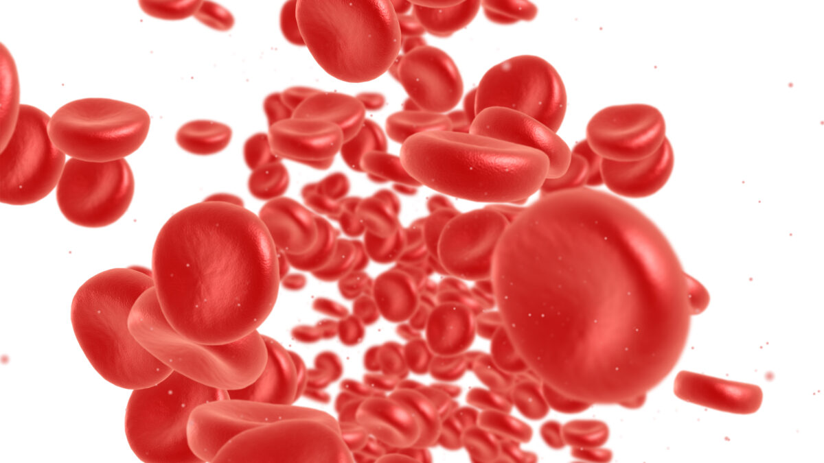
May 23, 2021Portal vein thrombosis (PVT) is a blood clot of the portal vein, also known as the hepatic portal vein. This vein allows blood to flow from the intestines to the liver. A PVT blocks this blood flow. Although PVT is treatable, it can be life-threatening.
The term portal venous thrombosis encompasses a broad spectrum of pathological conditions. Because the portal vein is narrowed or blocked, pressure in the portal vein increases.
This increased pressure (called portal hypertension) causes the spleen to enlarge (splenomegaly). It also results in dilated, twisted (varicose) veins in the oesophagus and often in the stomach. These veins can bleed profusely.
Which ultrasound scanner is best for portal vein thrombosis diagnosis?
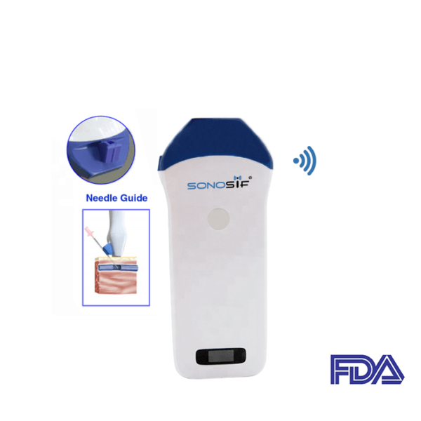
Using a high-frequency linear transducer is suitable for a PVT diagnosis. Based on this, the Mini-linear Handheld Wi-Fi Ultrasound Scanner MLCD is highly recommended to our gastroenterologist and hepatologist clients.
While regular ultrasounds use sound waves to produce images, they cannot show blood flow. Doppler ultrasounds, on the other hand, can use imaging to display blood circulation within the vessels. This can be used to diagnose portal vein thrombosis and determine how severe it is.
Acute thrombosis may be difficult to detect with grey-scale imaging alone, as the thrombus may be hypoechoic. With time, it becomes more echogenic and easier to identify. Colour Doppler should be able to demonstrate absent flow in the portal vein and even to detect partial thrombosis, but attention to the Doppler gain and filters is necessary to avoid color overwrite of partial thrombosis.
Moreover, Colour Doppler is also useful to help evaluate for tumour thrombus, which will show internal color vascularity. A bland thrombus, in comparison, is avascular on color Doppler.
The sonographic diagnosis of portal vein thrombosis is based on demonstration of echogenic material that obstructs the lumen of the vessel and the complete or partial absence of flow in the portal vein or on the presence of collateral circuits that by-pass the obstructed vessel, the most typical form being the Cavernoma, a tangle of tortuous vessels, irregular in calibre, that includes the vasa vasorum of the portal vein and the pericholecystic vessels.
References: Portal Vein Thrombosis, Portal Vein Thrombosis, PVT: The role of imaging,
Disclaimer: Although the information we provide is used by different doctors and medical staff to perform their procedures and clinical applications, the information contained in this article is for consideration only. SONOSIF is not responsible neither for the misuse of the device nor for the wrong or random generalizability of the device in all clinical applications or procedures mentioned in our articles. Users must have the proper training and skills to perform the procedure with each ultrasound scanner device.
The products mentioned in this article are only for sale to medical staff (doctors, nurses, certified practitioners, etc.) or to private users assisted by or under the supervision of a medical professional.


