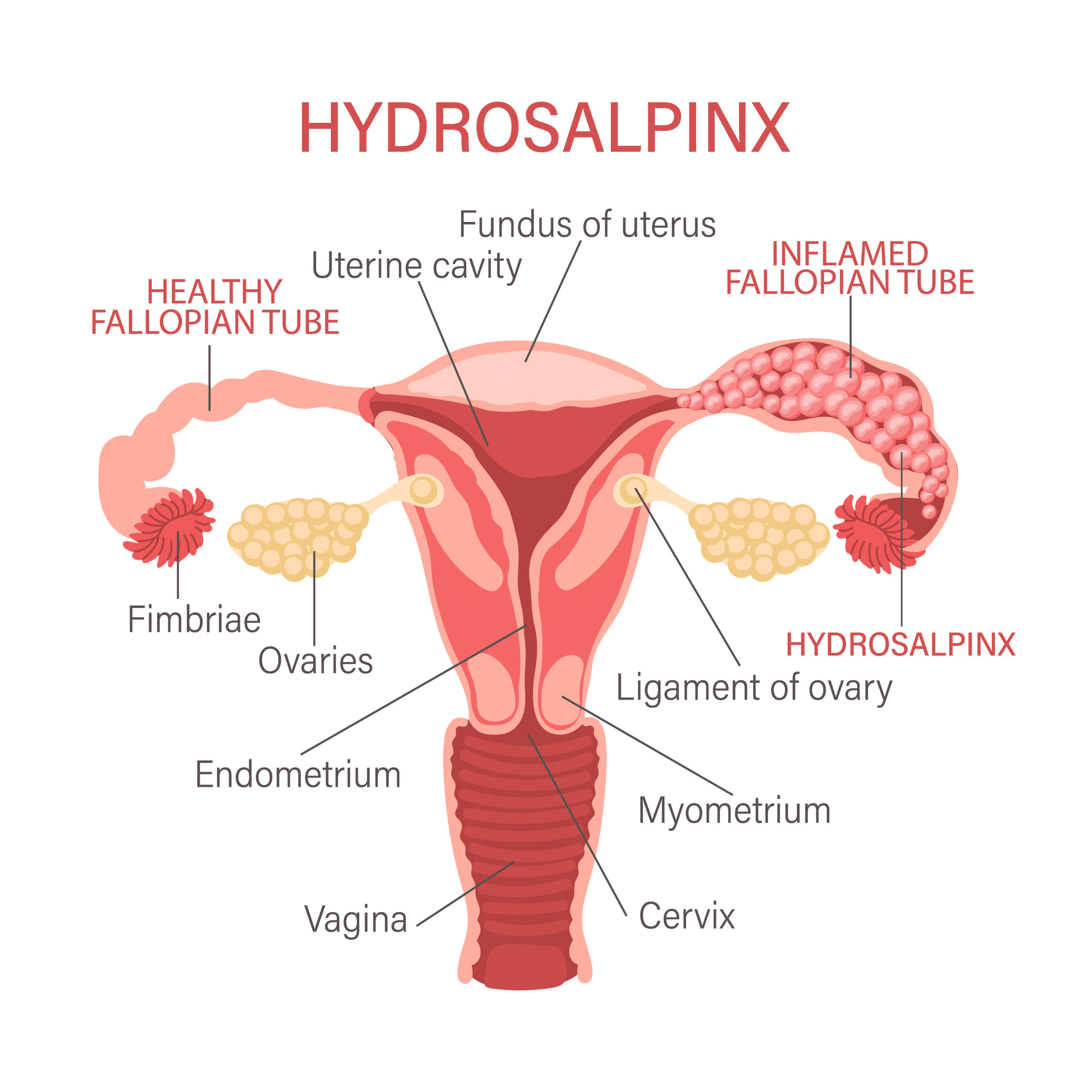
Hydrosalpinx Ultrasound
October 2, 2020
Speed and Direction of Blood flow – Doppler Ultrasound
October 3, 2020A radial arterial line (also an art-line or a-line) is a thin catheter inserted into an artery used in very ill or injured patients. It is most commonly used in intensive care medicine and anesthesia to monitor blood pressure directly and in real-time (rather than by intermittent and indirect measurement) and to obtain samples for arterial blood gas analysis.
The traditional radial artery cannulation can be challenging. There are many complications associated with it including temporary vascular occlusion, catheter-related infection. Therefore, this procedure must be done with great precision.
Ultrasound guidance has emerged as a valuable adjunct for the placement of radial artery catheters. The advantages of ultrasound guidance include real-time visualization of landmarks, improved pre-procedure planning, reduction in complications, less time spent at the bedside, and improved first-attempt success rates.
Which ultrasound scanner is best for radial arterial line placement?
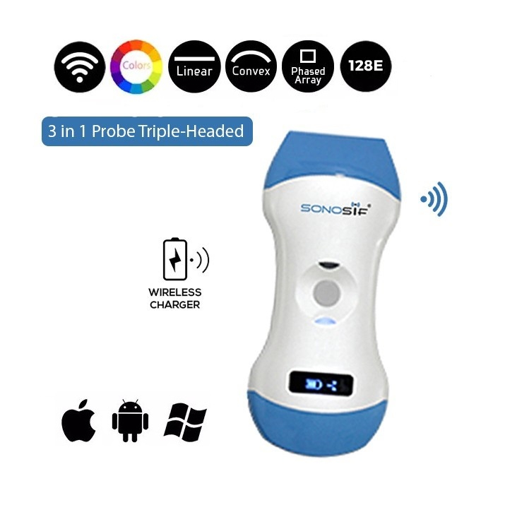
Physicians, Acute Care Nurse Practitioners (ACNP), ICU Physician Assistants(PAs), Anesthesiologist Assistants (CAAs), Nurse Anesthetists (CRNAs), and Respiratory Therapists will need the Color Doppler 3 in 1 Wireless Ultrasound Scanner 3in1-CLC1CD for this procedure.
The traditional method for radial artery catheter placement is to locate the vessel via palpation of the pulse or anatomic landmarks. Unfortunately, anatomic landmarks may not locate the radial artery in patients with severe hypotension, morbid obesity, and atherosclerosis. The radial pulse is weak or absent in these patients, making it difficult to locate the artery via palpation.
Other difficulties commonly encountered during radial artery catheter placement include the inability to thread the wire, hematoma formation, scarring from previous arterial catheters, atherosclerosis, and arterial spasm.
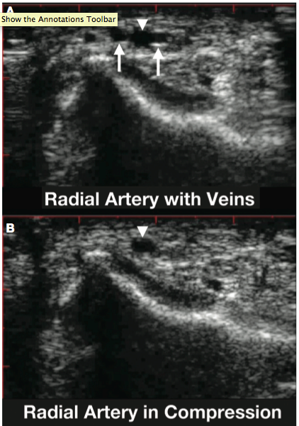
An ultrasound scanner with a Doppler mode can evaluate blood flow to aid in differentiation. Its linear or vascular probe can improve vessel visualization by adjusting the depth on the ultrasound to the minimum depth and adjusting the gain.
There are three primary techniques used for ultrasound-guided radial arterial line placement.
The first technique is the transverse method, where the probe is placed perpendicular to the artery. The skin is punctured at a 90° angle to the point on the probe that corresponds to the guide in the center of the screen. The needle will enter the screen from the center if properly oriented. By using real-time guidance, the needle can be redirected toward the vessel if the vessel is missed on the first pass.
The second technique is the longitudinal method, where the probe is oriented parallel to the vessel where it appears as a darkened area on the screen. The skin is punctured parallel to the probe in the exact center of the probe. The needle will enter the screen from either the left or right, depending on the probe’s orientation, and it will be visible entering the vessel when it reaches the correct depth. In some cases, the needle may lie parallel to the artery but may appear to have penetrated it on ultrasound images, in which case no blood return will be seen within the catheter chamber. In this case, the needle should be retracted, and the probe and catheter should be re-centered on the long axis of the artery, followed by reinsertion of the catheter.
The third method is the static technique, where the vessel is identified via ultrasound, and a mark is made on the skin along the artery’s path with a sterile marker.
Ultrasound guidance has been shown to reduce complications during central venous catheter insertion. The device also reduces the number of attempts required for a successful procedure and the likelihood of hitting the nerve bundle and other structures near the artery.
CLC1CD is a light-weighted portable ultrasound machine that is easy to use. It is iOS and Android compatible to give superior image quality. In addition, it is a wireless device, therefore; it can be used without worrying about fixing cables.

References: Respiratory Care. Review of Ultrasound-Guided Radial Artery Catheter Placement PDF


