- Immediate contact :
- +1-323-988-5889
- info@sonosif.com

Hysterosonography
October 8, 2020
Ultrasound-guided Vascular Cannulation
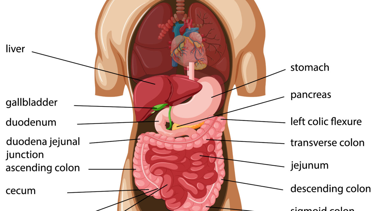
October 9, 2020Abdominal fascia refers to the various types of fascia found in the abdominal region. The superficial fascia of the abdomen consists (over the greater part of the abdominal wall) of a single layer containing a variable amount of fat; but near the groin, it is easily divisible into two layers, between which are found the superficial vessels and nerves and the superficial inguinal lymph glands.
Which Ultrasound Scanner is best for The Superficial Fascia of The Abdomen assessment?
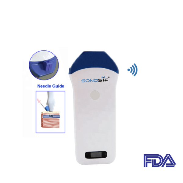
Using Ultrasound with a high-resolution and high-frequency transducer Mini-Linear Handheld Wi-Fi Ultrasound Scanner MLCD is strongly recommended to our Orthopedist clients. As a high-frequency probe offer superficial images, which is where The Superficial Fascia of The Abdomen is.
Using ultrasonography, fibrous connective tissue layers appear as echogenic and echo-lucent bands. Ultrasonography has the biggest advantage of assessing the sliding between the various fascial layers in real-time and permitting the accurate measurement of their thickness.
The superficial fascial system is highly abundant in some patients but has a limited presence in others. Individual-specimen mean gray value and whole-patient mean gray value positively correlated with tissue tensile strength (p = 0.006) and patient-average tissue tensile strength (p = 0.036), respectively. Whole-patient means gray value accounted for 98.5 percent of the variance seen in patient-average tensile strength, making it a strong predictor for tensile strength.
Moreover, for a healthy person, doctors need to position The probe along the right lateral abdominal wall at the height of the 12th rib: above the umbilicus at the linea alba, to the side of and approximately 2 cm from the umbilicus, along the mammillary line and the anterior axillary line. Each rater should measure about 17 anatomical structures six times during two sessions. Doctors admit that the reliability of using ultrasound in this procedure is good under both conditions for the fasciae and excellent for the muscles.
To conclude, Portable ultrasound and image-processing technology can visualize, quantify, and predict subcutaneous tissue strength of the superficial fascial system. The superficial fascial system quantity correlates with suture tensile strength.
References: Abdominal wall, Ultrasound imaging of the Superficial Fascia of the Abdomen, The Muscles and Fasciæ of the Abdomen,
Disclaimer: Although the information we provide is used by different doctors and medical staff to perform their procedures and clinical applications, the information contained in this article is for consideration only. SONOSIF is not responsible neither for the misuse of the device nor for the wrong or random generalizability of the device in all clinical applications or procedures mentioned in our articles. Users must have the proper training and skills to perform the procedure with each ultrasound scanner device.
The products mentioned in this article are only for sale to medical staff (doctors, nurses, certified practitioners, etc.) or to private users assisted by or under the supervision of a medical professional.


