- Immediate contact :
- +1-323-988-5889
- info@sonosif.com

M-Mode Ultrasound Imaging for Identifying a Pneumothorax
June 10, 2022
The Use of Ultrasound Scanners in Medical Routine in Operating Room
August 15, 2022Intramuscular fat builds up both inside (intramyocellular) and outside (extramyocellular) muscle fibers. Healthy muscle includes around 1.5 percent intramyocellular fat, which can rise to more than 5 percent in obese people.
Indded, Intermuscular fat is the largest definition of fatty infiltration in muscle, referring to lipid storage in adipocytes underneath the deep fascia of muscle.
Increased intramuscular fat levels have been discovered in both obese insulin resistant patients and highly trained endurance athletes; these contradictory findings have led to the conclusion that lipids accumulated inside muscle cells are not necessarily damaging to the cell.
An ultrasound scanner is essential for aiding in the assessment of intramuscular fat in muscles.
Actually, muscle ultrasonography is a feasible and reproducible imaging tool for determining the percentage of intramuscular fat. It is emerging as an imaging technique for measurement of muscle quality.
The echogenicity of lean muscle tissue is minimal, but that of intramuscular fat and connective tissue is high. This approach uses gray scale analysis to quantify overall muscle echo intensity, with the premise that the greater the mean pixel intensity of a muscle region of interest, the worse the muscle quality; that is, more intramuscular fat.
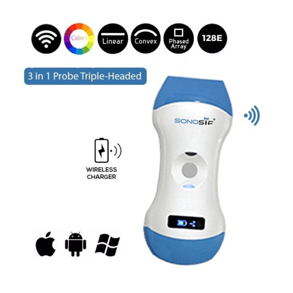
Muscle ultrasound, such as the Color Doppler 3 in 1 Wireless Ultrasound Scanner 3in1-CLC1CD, is a low-cost and easily accessible technique that may be used in those who cannot have other imaging technologies performed.
The 3 In 1 portable ultrasound CLC1CD provides qualitative and quantitative data. Thanks to the ultrasound machine’s smaller footprint, it aids in the evaluation of muscles. Further, this portable ultrasound is also a highly effective ultrasound scanner.
To sum up, ultrasound Scanner is a low cost, easily accessible, and highly reproducible method that can be an option for evaluating and Guiding Fat Gray Intramuscular.
References: Intermuscular Fat: A Review of the Consequences and Causes , Measurement of Intramuscular Fat by Muscle Echo Intensity
Disclaimer: Although the information we provide is used by different doctors and medical staff to perform their procedures and clinical applications, the information contained in this article is for consideration only. SONOSIF is not responsible neither for the misuse of the device nor for the wrong or random generalizability of the device in all clinical applications or procedures mentioned in our articles. Users must have the proper training and skills to perform the procedure with each ultrasound scanner device.
The products mentioned in this article are only for sale to medical staff (doctors, nurses, certified practitioners, etc.) or to private users assisted by or under the supervision of a medical professional.


