Pleural Drainage PLEFF
October 7, 2020
Hysterosonography
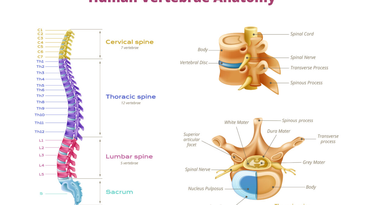
October 8, 2020Thoracic Paravertebral Block (TPVB) is the technique of injecting an anesthetic alongside the thoracic vertebra close to where the spinal nerves emerge from the intervertebral foramen. This produces unilateral, segmental, somatic, and sympathetic nerve blockage, which is effective for anesthesia and in treating acute and chronic pain of unilateral origin from the chest and abdomen.
The conventional technique of TPVB involves inserting the needle perpendicular to all planes, contacting the transverse process, and then walking off it with the needle. The commonly-used endpoint for needle insertion includes loss-of-resistance to air or saline, advancing a predetermined distance, or neurostimulation.
Ultrasound has been used to enhance the safety and efficacy of TPVB by determining the location and depth of the transverse process and the parietal pleura.
Which Ultrasound is best for Thoracic Paravertebral Block?
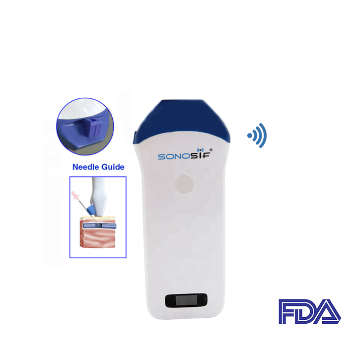
Using a high-frequency Transducer from 10 to 14MHz is the best for regional Ultrasound-guided blocks. Based upon, the Mini Linear Handheld Wi-Fi Ultrasound-Scanner MLCD is highly recommended to our Anesthesiologist clients.
The ultrasound transducer can be oriented in a transversal or sagittal direction, which results in a completely different representation of the TPV space on the ultrasound image. Bony structures, that is the rib, transverse process, and inferior articular process, constitute the main anatomical landmarks on the ultrasound.
Generally, “lateral” approaches are aimed close to the tip of the transversal process or in-between ribs, while “medial” techniques are performed medial to the costotransverse joint. The approaches are furthermore characterized by the usage of either an in-plane or out-of-plane technique and the direction of angulation in either the transversal or the sagittal plane.
For ultrasound examinations in the transversal plane, the transducer is placed lateral to the spinous process. Tilting and/or sliding the transducer in a craniocaudal direction generates three characteristic views of the TPV space. As for ultrasound examination in a sagittal orientation, the ultrasound transducer is positioned at approximately 5 cm lateral to the midline and gradually shifted toward medial.
With ultrasound guidance, the distance to the TPV space and pleura can be determined before performing a TPV block, whereas the position of the needle and the injection itself can be monitored in real-time during the block. This should contribute to an increased success rate and safety profile of the technique.
References: Ultrasound-guided TPVB, Thoracic Paravertebral Block, Different Approaches To US-guided TPVB
Disclaimer: Although the information we provide is used by different doctors and medical staff to perform their procedures and clinical applications, the information contained in this article is for consideration only. SONOSIF is not responsible neither for the misuse of the device nor for the wrong or random generalizability of the device in all clinical applications or procedures mentioned in our articles. Users must have the proper training and skills to perform the procedure with each ultrasound scanner device.
The products mentioned in this article are only for sale to medical staff (doctors, nurses, certified practitioners, etc.) or to private users assisted by or under the supervision of a medical professional.


