
Abdominal Pelvic Scan
October 20, 2020
Thoracentesis
October 20, 2020
Ultrasound of the Transrectal prostate uses sound waves to produce pictures of a man’s prostate gland and to help diagnose symptoms such as difficulty urinating or an elevated blood test result. It’s also used to investigate a nodule found during a rectal exam, detect abnormalities, and determine whether the gland is enlarged. Ultrasound is safe, non-invasive, and does not use ionizing radiation.
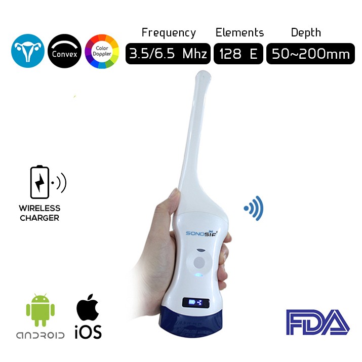
Which ultrasound Scanner is suitable for the Transrectal Prostate assessment?
Using the Convex and Transvaginal Color Doppler Double Head Wi-Fi Ultrasound Scanner CTC-3.1 is strongly suggested to our urologist clients. In which, the Transvaginal probe can provide images at different angles to help the doctor estimate the size of the patient’s prostate and spot abnormal growths.
It is also applied for the measurement of prostate volume, a study of echotexture, and illustration of tissue stiffness or elasticity. The Prostate Diagnosis provides images of the anterior fibromuscular stroma.
Transrectal Prostate Ultrasound can detect bacterial prostatitis appears as a hypoechoic rim around the prostate and shows an increased flow. Ultrasound is the primary tool in investigating a nodule, detect abnormalities, and determine whether the gland is enlarged.
Because ultrasound provides real-time images, it also can be used to guide procedures such as needle biopsies, in which a needle is used to sample cells (tissue) from an abnormal area in the prostate gland for later laboratory testing.
A prostate biopsy uses transrectal ultrasound imaging to guide several small needles through the rectum wall into areas of the prostate where the doctor sees something unusual.
The needles remove a tiny amount of tissue. Most doctors take six or more biopsies to test various areas of the prostate.
The scanning begins in the axial plane. The seminal vesicles are examined initially. As the probe is angled caudally the base of the prostate is seen. Once the prostate is examined in its entirety in this plane the probe is turned 90degrees in a sagittal plane. The probe is angled from one side to the other.
To sum up, prostate ultrasound is more accurate than an X-ray. This is because the technician can see the images as the transducer moves through the rectum rather than having to take a snapshot and develop the images. Ultrasound tests are also safer than X-rays because they don’t produce any dangerous radiation.
It is also faster than the computed tomography (CT) test, which provides 3-D images of your prostate and the areas around it. While CT scans require more preparation and time for testing, and they don’t provide real-time images.
References: Prostate Ultrasound, What to Expect from a Prostate Ultrasound, Transrectal Prostate Ultrasound and Biopsy
Disclaimer: Although the information we provide is used by different doctors and medical staff to perform their procedures and clinical applications, the information contained in this article is for consideration only. SONOSIF is not responsible neither for the misuse of the device nor for the wrong or random generalizability of the device in all clinical applications or procedures mentioned in our articles. Users must have the proper training and skills to perform the procedure with each ultrasound scanner device.
The products mentioned in this article are only for sale to medical staff (doctors, nurses, certified practitioners, etc.) or to private users assisted by or under the supervision of a medical professional.


