- Immediate contact :
- +1-323-988-5889
- info@sonosif.com

FAST: Focused Assessment with Sonography in Trauma
October 23, 2020
CVC: Central Venous Catheter
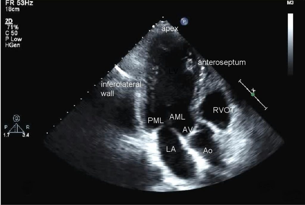
October 25, 2020Transthoracic Echocardiogram (TTE) is the most common non-invasive type of echocardiogram, which uses high-frequency Ultrasound to create a moving picture of the heart through the chest wall.
It is a test used to examine suspected problems with the valves or chambers of the heart, as well as the heart’s ability to pump blood. Yet, it is vital to identify potential causes of stroke.
This test may also be used to estimate the amount of blood pumped out of the left ventricle with each heartbeat, evaluate heart size and heart valve function, Identify areas of poor blood flow in the heart and areas of the heart muscle that are not contracting normally, and examine the previous injury to the heart muscle caused by impaired blood flow or evidence of congestive heart failure.
Which Ultrasound Scanner is suitable for Transthoracic Echocardiogram?
The sound waves used to obtain cardiac images in echocardiography are ultrasound waves that range in frequency from 2-12 MHz. As a result, adults are usually imaged using a 2-4 MHz transducer, while pediatric patients are imaged using a 7-12 MHz transducer. For this reason, the Convex and Linear Color Doppler Wi-Fi Double Head Ultrasound Scanner CLCD is highly suggested to our cardiologist clients.
It provides real time imaging of all four chambers and all four valves. Other visible structures on TTE include the aorta, the pericardium, pleural effusions, ascites, and inferior vena cava.
The ultrasound device can detect weakness of the heart, cardiac tumors and a variety of other findings can be diagnosed with a TTE.
Transthoracic echocardiography obtains cardiac images by placing the transducer head in contact with the thorax at one of the following locations, or acoustic windows: parasternal, apical, subcostal, and the suprasternal notch.

With advanced measurements of the movement of the tissue with time, ultrasound can measure diastolic function, fluid status, and ventricular dyssynchrony.

References: Transthoracic Echocardiogram, Echocardiogram,
Disclaimer: Although the information we provide is used by different doctors and medical staff to perform their procedures and clinical applications, the information contained in this article is for consideration only. SONOSIF is not responsible neither for the misuse of the device nor for the wrong or random generalizability of the device in all clinical applications or procedures mentioned in our articles. Users must have the proper training and skills to perform the procedure with each ultrasound scanner device.
The products mentioned in this article are only for sale to medical staff (doctors, nurses, certified practitioners, etc.) or to private users assisted by or under the supervision of a medical professional.


