- Immediate contact :
- +1-323-988-5889
- info@sonosif.com

The Use of Ultrasound in Hospice and Palliative Care
January 17, 2021
Post-Void Bladder
February 12, 2021To measure the direction and speed of a moving subject, Doppler ultrasonography is used. This is most generally applicable to the flow of blood in veterinary medicine.
The ultrasound wave reflected from the stationary object returns to the transducer at the same frequency at which it was transmitted when conducting ultrasound tests. However, if an ultrasound pulse is reflected from a moving target, the Doppler effect will change the frequency of the returned echo.
This variation in frequency is known as a Doppler Shift and can be sensed and displayed as color pixels or in graphical format by an ultrasound system.
More generally, Doppler ultrasound is used in echocardiographic exams. Areas of irregular or turbulent blood flow associated with heart changes can be visualized by mapping the rate and direction of blood flow. Furthermore, via the modified Bernoulli equation, the quantification of blood velocity in the heart chambers provides details on changes in blood pressure.
Which Ultrasound scanner is more suitable for Vascular access in newborn piglets?
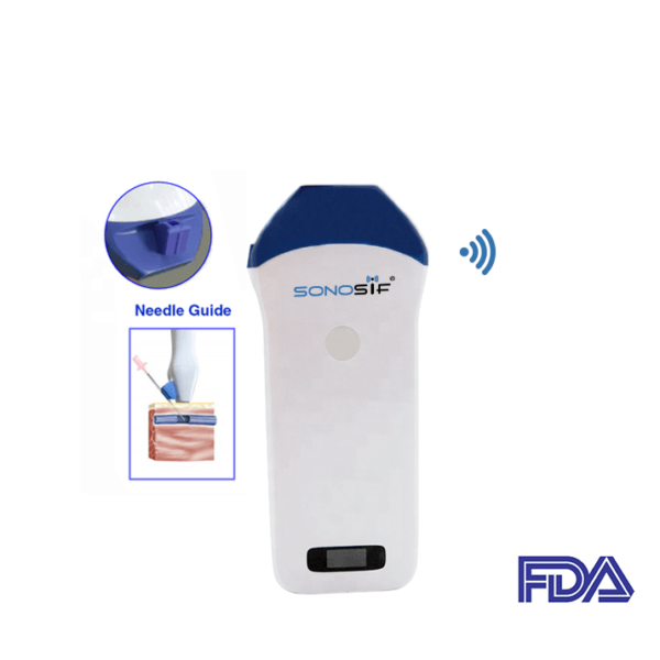
In newborn animals and particularly pigs, the use of a high-frequency ultrasound scanner with a needle guide is more appropriate for vascular entry.
For instance, the Mini Linear Handheld WiFi Ultrasound Scanner MLCD provides guided injections. This allows the practitioner to visualize the needle in real-time as it enters the body and traverses to the desired location.
Even in the most skilled hands, blind (injections without imaging) injections are not 100% effective and, in some joints, the accuracy is as poor as 30%-40%. The accuracy of almost any joint injection reaches 90 percent with ultrasound guidance and is reaching 100 percent in many cases.
In particular, the U.S. puncture instruction in real-time helps the vet to change the patent of the vessel and the location of the needle, catheter, and wire in the vessel. Ultrasound can also be used to assess the patient’s condition changes and thus the lumen cross-section based on the patient’s position change.
Using Color Doppler ultrasonic scanners with a needle guide increases needle placement and injection precision. Provides control for the needle penetration direction, needle depth, or angles without the possibility of disrupting neighboring designs and decreases the time of intra-vascular entry.
In brief, Ultrasound guidance is a valuable method to help locate venous and arterial arteries and can lead to rapid and reproducible venepuncture and vascular access in piglets.
References: Ultrasound Doppler explained for vets, Color Doppler Ultrasound Assessment,
Disclaimer: Although the information we provide is used by different doctors and medical staff to perform their procedures and clinical applications, the information contained in this article is for consideration only. SONOSIF is not responsible neither for the misuse of the device nor for the wrong or random generalizability of the device in all clinical applications or procedures mentioned in our articles. Users must have the proper training and skills to perform the procedure with each ultrasound scanner device.
The products mentioned in this article are only for sale to medical staff (doctors, nurses, certified practitioners, etc.) or to private users assisted by or under the supervision of a medical professional.


