Confirmation of Nasogastric Tube Placement
October 6, 2020
Traumatic Extremity Injuries
October 7, 2020A lumbar Puncture which is also known as a spinal tap is performed in the lower back in the lumbar region. During a lumbar puncture, a needle is inserted between two lumbar bones (vertebrae) to remove a sample of cerebrospinal fluid ( it’s the fluid that surrounds the brain and spinal cord to protect them from injury).
It can help diagnose serious infections, such as meningitis; other disorders of the central nervous system, or cancers of the brain or spinal cord. Moreover, Lumbar Puncture is used to inject anesthetic medications or chemotherapy drugs into the cerebrospinal fluid.
Ultrasound mapping of the Lumbar Puncture reveals anatomical that is not obtainable by physical examination, including depth of the ligamentum flavum, width of the interspinous spaces, and spinal bone abnormalities.
Which ultrasound is best for Lumbar Puncture imaging?
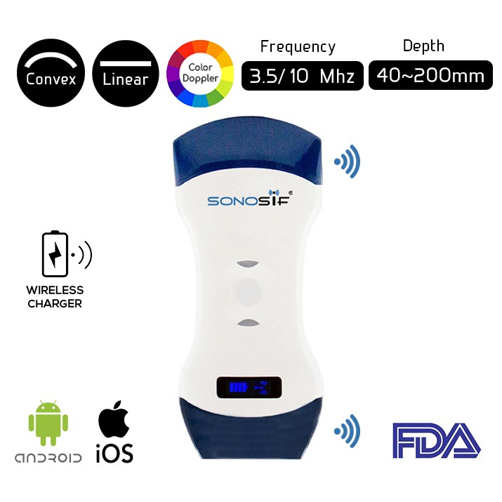
For Lumbar Puncture, a high-frequency linear array transducer and curvilinear transducer are useful and convenient. Yet, the first one generates high-resolution images and is preferred in lean patients and for training novice providers. While the second provides deeper penetration to visualize the spinal structures in overweight and obese patients. For this reason, our SONOSIF ‘s Research and Development team highly recommends the Convex and Linear Color Doppler Wi-Fi Double Head Ultrasound Scanner CLCD to our Neurologist clients.
When ultrasound guidance is available, along with providers who are appropriately trained to use it, Ultrasound guidance should be used for site selection of LPs to reduce the number of needle insertion attempts and needle redirections and increase the overall procedure success rates, especially in patients who are obese or have difficult-to-palpate landmarks.
The ultrasound transducer must be placed in a sterile plastic sheath when using real-time guidance. After entering the transducer over the widest lumbar interspinous space, rotate the transducer 45° toward the midline into an oblique paramedian view.
The transducer is aligned in a plane from the spinous process of the superior vertebra to the lamina of the inferior vertebra. The lamina, ligamentum flavum, spinal canal, and posterior longitudinal ligament are visualized. Slide the transducer in the same plane 1 to2 cm craniomedially to ease the insertion of the needle underneath the transducer. The spinal needle is inserted in the plane of the ultrasound beam.
To conclude, Ultrasound guidance for Lumbar Puncture improves success rates, decreases needle redirections and traumatic taps, and can decrease total procedure time. When static ultrasound guidance is used, visualization of the spinous processes is the key element to mark the needle insertion site. When real-time ultrasound guidance is used, the needle is inserted under direct visualization using a paramedian approach.
References: Lumbar Puncture, Ultrasound guidance to Lumbar Puncture, US-guidance to LP
Disclaimer: Although the information we provide is used by different doctors and medical staff to perform their procedures and clinical applications, the information contained in this article is for consideration only. SONOSIF is not responsible neither for the misuse of the device nor for the wrong or random generalizability of the device in all clinical applications or procedures mentioned in our articles. Users must have the proper training and skills to perform the procedure with each ultrasound scanner device.
The products mentioned in this article are only for sale to medical staff (doctors, nurses, certified practitioners, etc.) or to private users assisted by or under the supervision of a medical professional.


