Ultrasound-guided Breast Reduction
August 5, 2021Ultrasound-guided Male Breast (Gynecomastia) Surgery
August 23, 2021The medical term for a breast lift is mastopexy. A cosmetic surgeon lifts and reshapes the breasts to give them a firmer, rounder appearance in this surgery. The procedure also lowers the size of the areola, which is the colourful circle surrounding the nipple and removes excess skin around the breast.
This can assist a patient’s look by restoring her youthful, feminine proportions and making bras and swimsuits fit more comfortably and attractively.
Large, stretched areolae can also be decreased during breast lift surgery, resulting in a more proportionate, natural-looking breast.
Ultrasound is used to evaluate breast tissue and determine if patients are surgical candidates. When done by a technically qualified surgeon, ultrasound is a safe and effective method of mastopexy in selected patients.
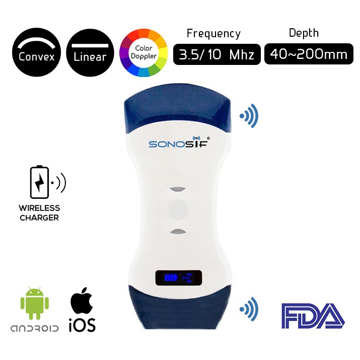
For example, the Convex and Linear Color Doppler WiFi Double Head Ultrasound Scanner CLCD enables the practitioner to overcome obstacles and any associated problems, as well as the diagnosis and follow-up of further breast lesions.
Thanks to two parts, it is more convenient and cost-effective than purchasing two separate single-headed probes. The doctor can assess the more superficial portions of the body using the Linear side of the Doppler, while the Convex side is utilized for in-depth exams.
The Double Head Ultrasound Linear side Frequency goes from 40 to 100 mm. Its frequency varies from 7.5 to 10 MHz. The linear probe is usually used to visualize, Breast, and vascular applications.
Moreover, The Color Double Head WiFi Ultrasound Scanner’s Convex probe offers a frequency range of 3.5 to 5 MHz. It has a depth range of 90 to 305 mm, allowing for improved monitoring, examination, and diagnosis.
The surgeon can use an ultrasound device to evaluate the morphology, form, and content of the breasts, as well as monitor the tissues and axillae. Furthermore, if the patient wishes, ultrasonography in the breast implant may be required later.
References: What is breast lift surgery?, Mastopexy
Disclaimer: Although the information we provide is used by different doctors and medical staff to perform their procedures and clinical applications, the information contained in this article is for consideration only. SONOSIF is not responsible neither for the misuse of the device nor for the wrong or random generalizability of the device in all clinical applications or procedures mentioned in our articles. Users must have the proper training and skills to perform the procedure with each ultrasound scanner device.
The products mentioned in this article are only for sale to medical staff (doctors, nurses, certified practitioners, etc.) or to private users assisted by or under the supervision of a medical professional.


