Tendons of the medial and lateral compartment of the ankle
April 21, 2021
Ultrasound-Guided Anserine bursa
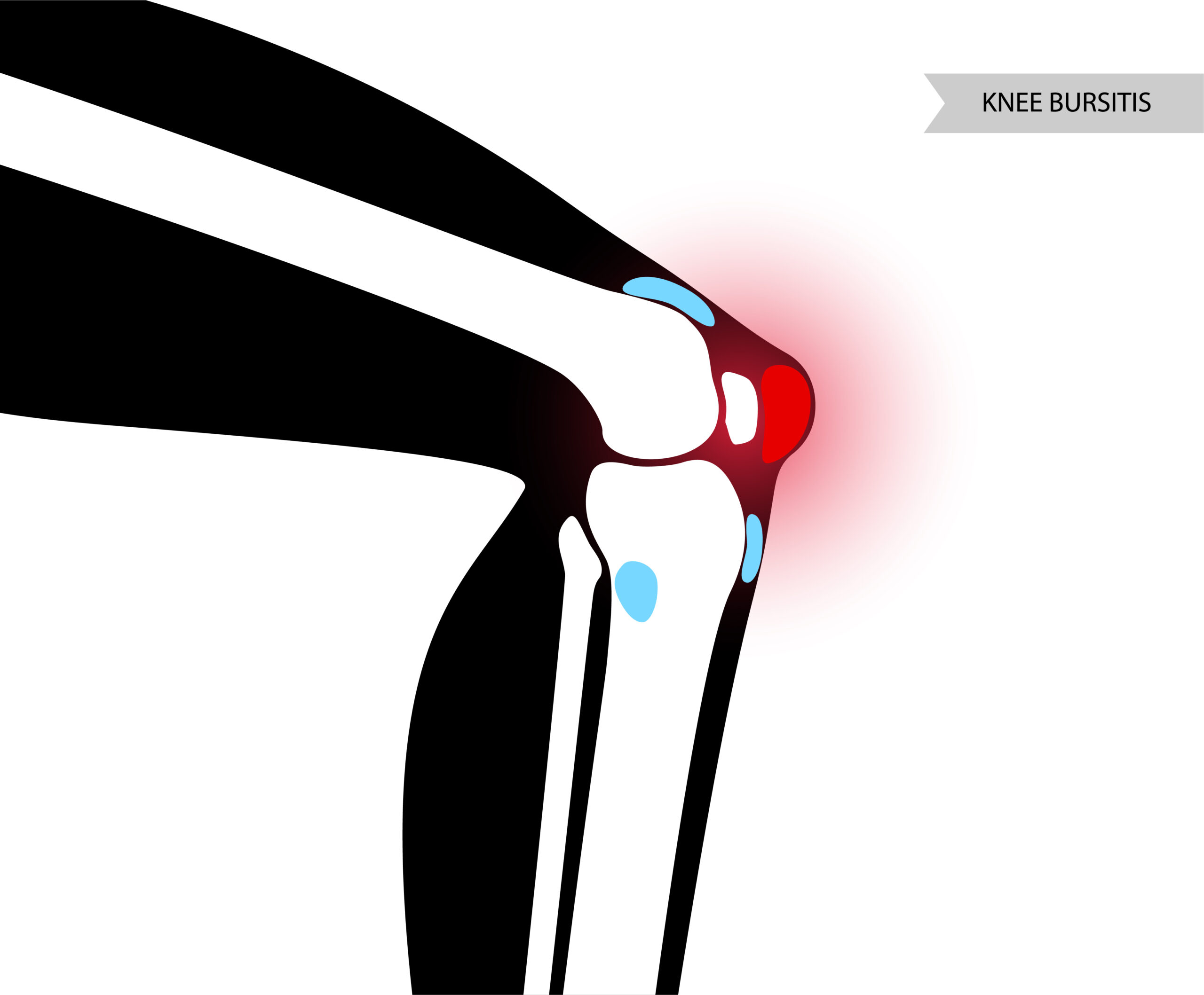
May 6, 2021Suprapatellar recess can be found just above the knee. It’s located between the femur (thigh bone) and the quadriceps tendon.
It’s also known as the suprapatellar bursa or suprapatellar pouch. Suprapatellar recess is located proximal to the knee joint, between the femoral and suprapatellar fat pads. As with all bursae, its purpose is to reduce friction between moving structures.
Inflammatory joint conditions most commonly involve the knee. In certain studies, ultrasonography is similar to clinical evaluation in determining the presence and position of knee joint effusion. Soft tissue abnormalities (suprapatellar bursitis, knee effusion, or Baker’s cyst) were observed by ultrasonography at 54/130 (42 percent) of the locations.
Which Ultrasound Scanner is the best for Suprapatellar recess assessment?
During the evaluation of the suprapatellar bursa, a high-frequency linear transducer with a range of 5 to 12 MHz is needed. Thereby, our orthopedist clients tend to use either the USB Linear 5-12MHz Ultrasound Scanner USB-UL2.
USB-UL2 plays an important role in improving point-of-care, clinical diagnostic methods and efficiency of clinic diagnosis. With its sealed head and its USB connector, a steady ultrasound signal makes the signal transmission faster, and it provides a high-quality image that guides the physician to clear decisions during the assessment.
Under ultrasound guidance, the suprapatellar approach avoids any tendons or ossic or ligament structures and facilitates the provider for easy and accurate arthrocentesis.
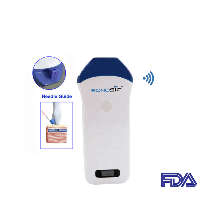
The Mini Linear Handheld WiFi Ultrasound Scanner MLCD might be used also by physicians for both the identification of the effusion and needle guidance for the arthrocentesis.
It comes with a needle guide holder. Hence, it can be directly set to the guide pin frame. Coupled with the software that can quickly locate the depth and diameter of puncture’s navigation.
MLCD aids in the reduction of attempts and the improvement of procedural trust in the hands of healthcare providers.
Ultrasonography is useful for detecting knee effusion and providing a real-time visualization technique for joint aspiration. It will also help with the diagnosis of a patient who has a sore, or hurting knee.
References: Suprapatellar Bursa, Suprapatellar Bursitis
Disclaimer: Although the information we provide is used by different doctors and medical staff to perform their procedures and clinical applications, the information contained in this article is for consideration only. SONOSIF is not responsible neither for the misuse of the device nor for the wrong or random generalizability of the device in all clinical applications or procedures mentioned in our articles. Users must have the proper training and skills to perform the procedure with each ultrasound scanner device.
The products mentioned in this article are only for sale to medical staff (doctors, nurses, certified practitioners, etc.) or to private users assisted by or under the supervision of a medical professional.


