
Ultrasound-guided Breast reduction
February 17, 2022
Vitreous Hemorrhage Ocular Ultrasound Diagnosis
February 17, 2022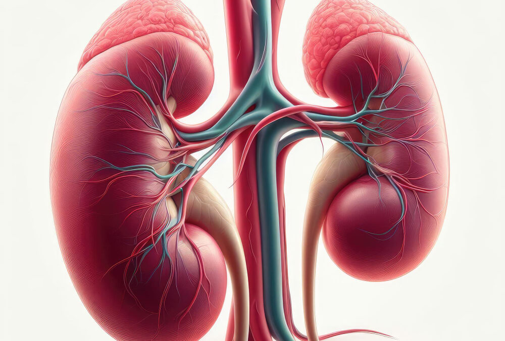
Each kidney in an adult is around 3 cm thick, 6 cm wide, and 12 cm long. It has a bean-shaped indentation on the medial side, known as the hilum.
The hilum connects to the renal sinus, a vast hollow within the kidney. The renal artery enters the kidney at the hilum, where the ureter and renal vein exit the kidney.
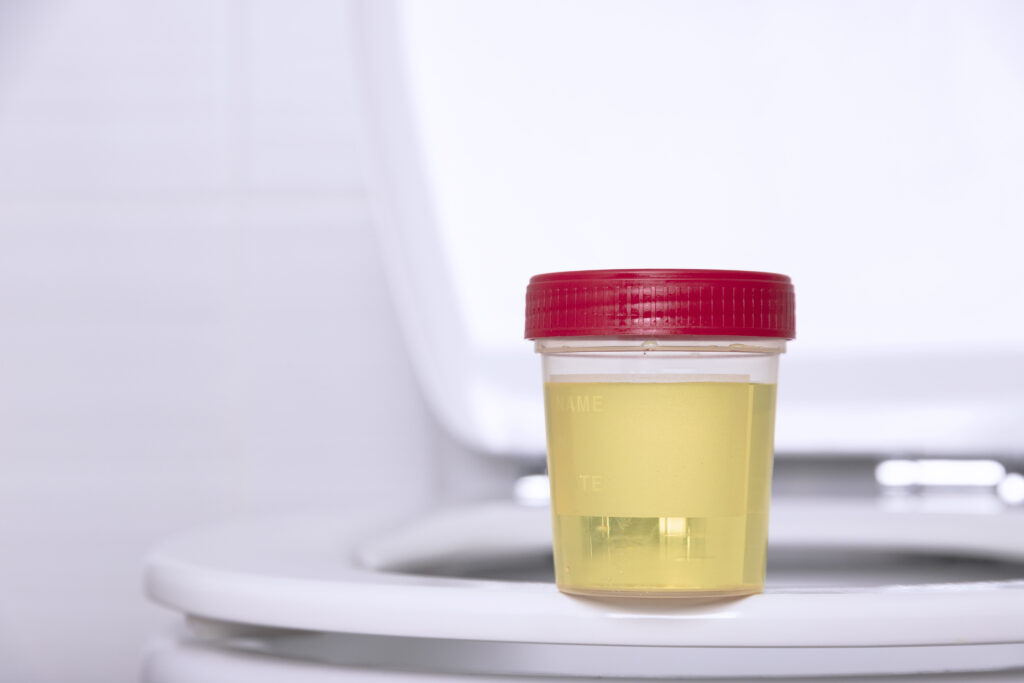
The renal pelvis, which is positioned in the renal sinus and connects to the ureter, is located in the central portion of the kidney. The renal pelvis is a vast cavity in which urine is collected when it is produced.
Ultrasound is a critical tool in the diagnosis and treatment of nephrological patients, allowing for the examination of the shape, cavities, and function of the kidneys.
Ultrasonography machines capable of completing studies in two-dimensional and Doppler modes are required for nephrology ultrasound investigations. A low-frequency convex transducer for abdominal cavity analysis (kidneys, bladder, prostate, liver-bile duct, and abdominal aorta) and a high-frequency linear transducer for investigation of more superficial tissues should be included in the equipment (pleura-lung, parathyroid glands, carotid, and femoral arteries).
The ultrasound gadget should also be capable of cannulating the jugular and femoral veins. For HD scanning, the same linear transducer will be used for puncture and investigation of vascular access.
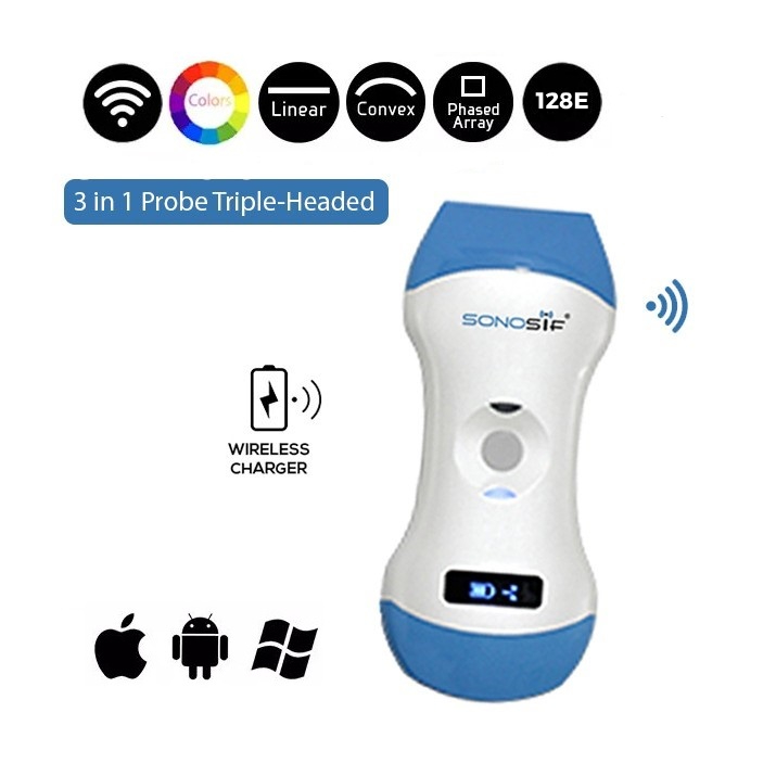
The Color Doppler 3 in 1 Wireless Ultrasound Scanner 3in1-CLC1CD is recommended to satisfy the ultrasound parameters required for various scanning procedures.
With a Convex 3.5/5MHz and Linear 7.5/10MHz, this ultrasound scanner is suited for both adult and pediatric patients. It also has a color doppler for determining the renal artery’s systolic velocity.
There is no need to change the probe’s head because the software can be changed instead. The engineers at SIFSOF designed this portable ultrasound machine specifically for nephrology ultrasonography diagnosis and intervention.
Real-time ultrasonography, as achieved by the 3in1-CLC1CD in the application of vascular access, means that the progression of the needle to the vessel is done under continuous visualization by this imaging approach, which has numerous advantages.
With all these advanced options, the 3in1-CLC1CD should be nephrologists’ first choice as it provides accurate scanning imagery following easy scanning procedures.
Reference: The role of ultrasound guidance for vascular access
Ultrasonography of the Kidney: A Pictorial Review


