- Immediate contact :
- +1-323-988-5889
- info@sonosif.com

Fetal Morphology Assessment: FMA
June 11, 2021
CP: Chronic Pain
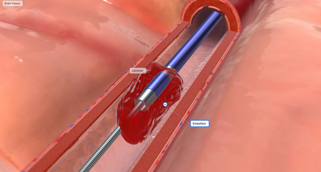
June 15, 2021A vascular access procedure involves the insertion of a flexible and sterile thin plastic tube, or catheter, into a blood vessel to provide an effective method of drawing blood or delivering medications, blood products, or nutrition into a patient’s bloodstream over a period of weeks, months or even years.
Indeed, Cannulation of veins and arteries is a crucial element of patient care for fluid and drug delivery as well as monitoring.
Ultrasound in Vascular Access improves the safety of Vascular Punctures. Not just by lowering the number of failed puncture attempts, but also by lowering the problems and expenses associated with this medical method.
Which ultrasound scanner do doctors use for Vascular Access?
Using a high-frequency Ultrasound Scanner reduces complications and vascular puncture costs as well as increasing the procedure’s success rate. Hence, it is vital equipment in the routine practice of this procedure.
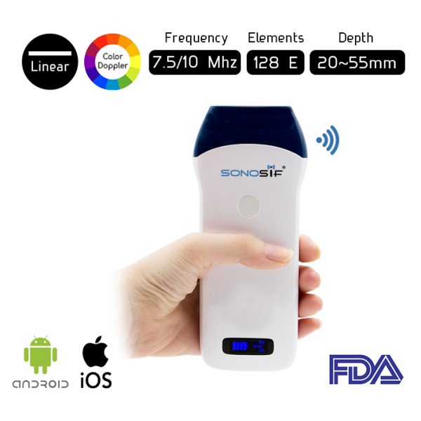
For instance, the Wireless Color Doppler Linear Ultrasound Scanner L2CD works in a way that the needle level touches the ultrasound beam in a pre-established depth. Which reduces catheter placement failure rates and the relative risk of mechanical complications.
L2CD is used to assess and identify a vein that is suitable for catheter placement. It also allows the interventional radiologist the ability to identify appropriate veins that may be larger and deeper than veins that can be seen or felt on the skin surface.
Ultrasound guiding for vascular access is most successful when utilized in real-time (during needle advancement) using a sterile method that includes sterile gel and sterile probe covers. The needle is shown on the imaging display and is concurrently steered toward the target vessel, away from neighboring structures, and advanced to an acceptable depth.
References: Vascular Access Ultrasound, Guidelines for Performing Ultrasound Guided Vascular Cannulation.


