The Superficial Fascia of The Abdomen
October 8, 2020
Ultrasound-guided Arthrocentesis
October 9, 2020Vascular Cannulation is common practice in critical care, and it is traditionally performed using the landmark technique, yet it is not exempt from failure and complications. In this regard, Ultrasound-guided vascular cannulation (USGVC) has been shown to improve the procedure success rate and reduce its associated complications.
Which Ultrasound is suitable for Vascular Cannulation?
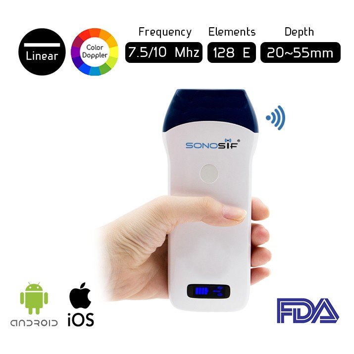
Since Vascular structures are superficial, a linear array transducer (frequency from 7 to 10) MHz is commonly used to recognize the vessels, with the selection of a preset corresponding to veins or arteries, as the case may be. For this reason, the Wireless Color Doppler Ultrasound Scanner L2CD is highly recommended to our Cardiologists clients.
Ultrasound-guided vascular cannulation is becoming the modality of choice to obtain a tool to obtain secure vascular access in critical care patients.
Ultrasound guidance for vascular access is most effective when used in real-time (during needle advancement) with a sterile technique that includes sterile gel and sterile probe covers. The needle is observed on the image display and simultaneously directed toward the target vessel, away from surrounding structures, and advanced to an appropriate depth. Static ultrasound imaging uses ultrasound imaging to identify the site of needle entry on the skin over the underlying vessel and offers the appeal of nonsterile imaging.
If ultrasound is used to mark the skin for subsequent cannulation without real-time (dynamic) use, ultrasound becomes a vessel locator technique that enhances external landmarks rather than a technique that guides the needle into the vessel. Both static and real-time ultrasound-guided approaches are superior to a traditional landmark-guided approach.
Ultrasound-guided vascular cannulation may lead inexperienced operators to use needle angle approaches that lead to an increased risk for complications. Traditional approaches and techniques mustn’t be abandoned with ultrasound guidance, particularly during cannulation of the SC vein, in which a steeper needle entry angle may lead to a pleural puncture. The needle is directed toward the sternal notch in the coronal plane.
To sum up, Ultrasound guidance can be used for placing central venous catheters as well as for placing peripheral venous catheters. Clinicians who place central venous access devices (occasionally or frequently) are strongly encouraged to learn ultrasound-guided techniques.
References: Ultrasound-guided Vascular Cannulation in critical care patients, Guidelines for performing Ultrasound-guided Vascular Cannulation,


