
Abscesses
October 14, 2020
Gastroesophageal Reflux Disease (GERD)
October 15, 2020
VeniPuncture is a procedure in which a needle is used to take blood from a vein, usually for laboratory testing. Venipuncture may also be done to remove extra red blood cells from the blood, to treat certain blood disorders. Also called blood draw and phlebotomy.
The use of ultrasound to guide catheter placement reduces the number of access attempts and may reduce other complications as well.
Which Ultrasound Scanner is suitable for VeniPuncture?
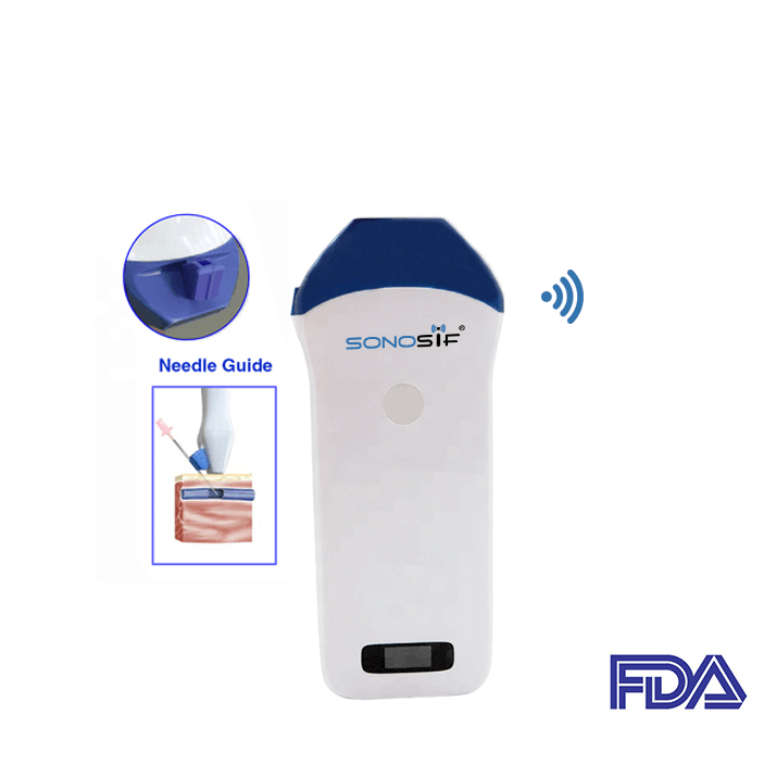
During this procedure, the practitioner needs a high-frequency Ultrasound Scanner from 10 to 14 MHz and high-image resolution, in order to confirm the course of blood flow in the vessels, also in aligning the axes of the echo probe and the VP needle. For this reason, the Mini-Linear WiFi Ultrasound Scanner MLCD is highly recommended to our phlebotomist clients.
An advantage of Mini-Linear WiFi Ultrasound Scanner MLCD guided procedures is the control of the needle insertion path and, if needed, the possibility to modify the needle depth or angle without the risk of damaging adjacent structures.
Since needle visualization is critical. The use of a needle guide keeps the needle aligned with the ultrasound probe’s sound beam, leading to Improved needle visualization, Reduced procedure time, and Confident clinical outcomes.
Depending on the positioning of the ultrasound probe, and image resolution, there will be an ultrasound-guided venipuncture in the short or long axis.
The one on the short axis is called “out of plane“, while the second one “in-plane“: the choice may depend on the diameter, on the depth of the vein, and on the practicality of the healthcare professional.
The ultrasound-guided VP should be preceded by a determination of the most appropriate vein for access, based upon systematic ultrasound evaluation of possible access sites. In addition, it is indicated not only for adult applications but also for pediatric and neonatal ones.
The VP should be performed using dynamic “real-time” ultrasound guidance, not “static” ultrasound localization of the vein with subsequent “blind” venipuncture.
Ultrasound-guided injections allow the practitioner to visualize the needle in real-time as it enters the body and traverses to the desired location; it provides more accurate needle placement in all the body joints. This assures that the medication is accurately injected at the intended site.
References: Ultrasound and VeniPuncture, Principles of ultrasound-guided venous access


