Interscalene Plexus Nerve Blocks (PNB)
October 13, 2020
Abscesses
October 14, 2020Venous Ultrasound is a technique that uses sound waves to produce images of the veins in the body. This procedure is commonly used to search for blood clots, especially in the veins of the leg – a condition often referred to as deep vein thrombosis.
In addition it aids in guiding placement of a needle or catheter into a vein. Sonography can help locate the exact site of the vein and avoid complications, such as bleeding or damage to a nearby nerve or artery. VU also helps map out the veins in the leg or arm so that pieces of vein may be removed and used to bypass a narrowed or blocked blood vessel.
Which Doppler Ultrasound is suitable for venous imaging?
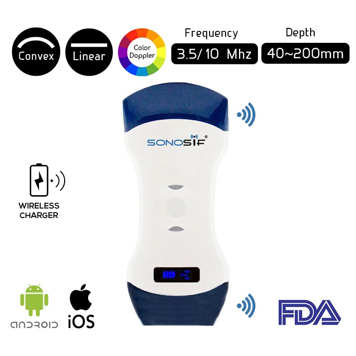
Using a 5 MHz US frequency is needed for vascular and a high-frequency US of 10 MHz is needed for venous ultrasound examination. Because it is the best option for detecting clots, aid in guiding placement of a needle or catheter into a vein, and map out the veins in the arm or leg, the Convex and Linear Color Doppler Wi-Fi Double Head Ultrasound Scanner CLCD is highly recommended to our Cardiologist and Radiologist clients.
Doppler ultrasound images can help the physician to see and evaluate not only blockages to blood flow (such as clots), narrowing of vessels, tumors and congenital vascular malformations, increased blood flow, which may be a sign of infection but also reduced or absent blood flow to various organs, such as the testes or ovary. Besides, it can help the Doctor see the reduced or absent blood flow to various organs or if it’s an increased blood flow, which is a sign of an infection.
The venous system of the lower extremities is divided into three groups: deep, superficial, and perforating veins. In US examination of the lower extremity veins, knowledge of the venous anatomy as well as appropriate patient positioning and transducer placement are important for optimal imaging and accurate diagnosis.
The benefits of this procedure is that it is safe and painless. It gives a clear picture of soft tissues and inner-organs without the danger of radiation. Yet, the Venous Doppler Exam does not require any needle injection or intervention, only the presence of a trained medical examiner and a High Resolution Ultrasound Scanner.

References: Ultrasound – Venous, Venous Doppler Ultrasound, Doppler Ultrasound Exam of Arm and Leg
Disclaimer: Although the information we provide is used by different doctors and medical staff to perform their procedures and clinical applications, the information contained in this article is for consideration only. SONOSIF is not responsible neither for the misuse of the device nor for the wrong or random generalizability of the device in all clinical applications or procedures mentioned in our articles. Users must have the proper training and skills to perform the procedure with each ultrasound scanner device.
The products mentioned in this article are only for sale to medical staff (doctors, nurses, certified practitioners, etc.) or to private users assisted by or under the supervision of a medical professional.


