US-Guided Cavity Analysis and Vascular Access
February 17, 2022
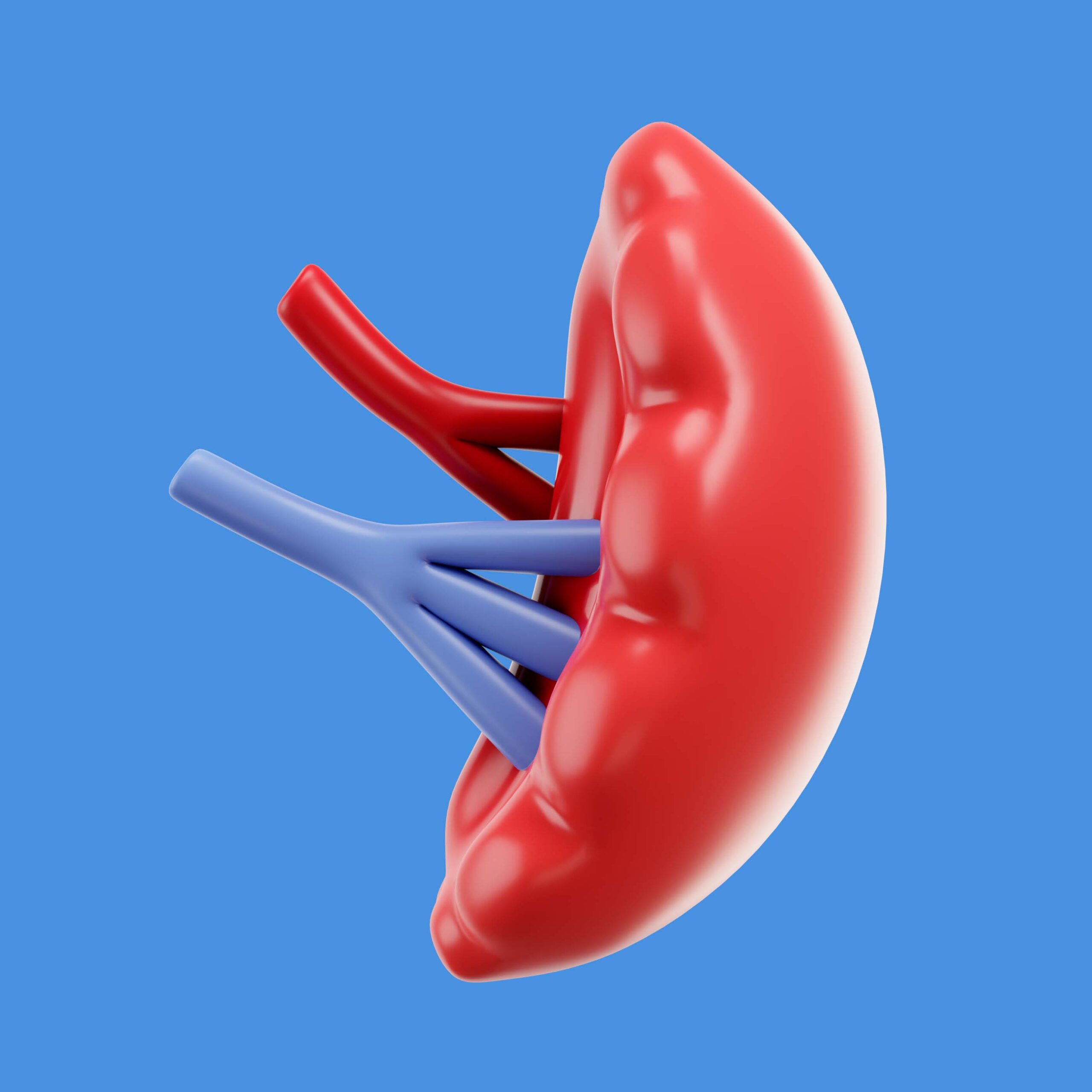
Ultrasound Scanner for Abnormal Enlargement of the Spleen
April 7, 2022Vitreous hemorrhage (VH) is a serious ocular ailment that can result in a sudden loss of visual acuity (VA) and is frequently a consequence of another condition.
Proliferative diabetic retinopathy (PDR), retinal vein occlusions (RVOs), ocular trauma, posterior vitreous detachment with or without a retinal tear, and other conditions are some of the most common causes of VH.
Proliferative retinopathy, a disorder in which new, aberrant blood vessels sprout on the surface of the retina, can cause a vitreous hemorrhage. The process is known as neovascularization. These new blood vessels may continue to grow and expand through the vitreous into the pupil area if they are not addressed. This can cause ocular pressure (pressure inside the eye) to rise, putting strain on the optic nerve.
Doctors will examine the patient’s eyes and analyze their medical history to establish the origin of the hemorrhage and provide therapeutic recommendations.
As per protocol, a 10 MHz B-scan ultrasonic probe (with a depth of exploration of 20 to 60 mm, focus of 21 to 25 mm, the axial resolution of 150 m, and lateral resolution of 300 m) is used to evaluate the temporal quadrant of the globe with the patient in the dorsal decubitus posture.
The optic nerve head, macula, peripheral retina, and external rectus muscle can all be seen in a longitudinal picture. As a result, for right eyes, the 9 o’clock meridian is examined, while for left eyes, the 3 o’clock meridian is examined.
The Ophthalmic Digital Ultrasound Scanner, A/B Scan, 15 inch LCD Screen Opthta2-CD is highly recommended for ocular ultrasound screening methods for Vitreous hemorrhage.
This ultrasound allows operators to readily view the front and posterior parts of the eye, providing crucial information that would otherwise be unavailable through clinical examination alone.
The Opthta2-CD has a B-scan with a frequency range of 10MHz/20MHz (optional), is magnetically operated and noiseless, and has a 60 mm depth.
This technology has shown to be a fantastic tool for detecting Vitreous hemorrhage. With a probe gain of 30dB-105dB, it improves the region of the vitreous body and retina that is ideal for grading vitreous hemorrhage.
The Opthta2-CD also has an A-scan mode for measuring anterior chamber depth, lens thickness, vitreous body length, and total length during cataract surgery to determine the best lens replacement and for tumor diagnosis.
Reference: Vitreous Hemorrhage: Diagnosis and Treatment
Scale for Photographic Grading of Vitreous Haze in Uveitis


