
Ultrasound-Guided Paracentesis
October 15, 2020
Hemodialysis Catheters
October 16, 2020
The abdominal wall surrounds the abdominal cavity, providing it with flexible coverage and protecting the internal organs from damage. It is bounded superiorly by the xiphoid process and costal margins, posteriorly by the vertebral column, and inferiorly by the pelvic bones and inguinal ligament.
Sonography is usually regarded as a first‐line imaging modality for masses and masslike lesions in the abdominal wall.
Which Ultrasound Scanner is suitable for abdominal wall imaging?
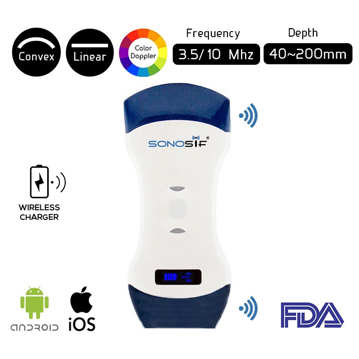
Using the Convex and Linear Color Doppler Wi-Fi Double Head Ultrasound Scanner CLCD is strongly suggested to our gastroenterologist clients. In which it helps the practitioner to decide carefully whether the discomfort is coming from the abdominal or the inside.
The High-frequency linear transducer of 7.5 to 10 MHz allows a detailed assessment of anatomy and high-quality imaging of superficial pathology. The exact location of lesions concerning the layers of the abdominal wall can be determined. While using the low-frequency convex transducer of 3.5 to 5 MHz is the best for transabdominal imaging.
Sonography has been reported to significantly improve the sensitivity and specificity for preoperatively diagnosing several lesions, such as lipoma and an epidermoid cyst.
Many illnesses can compromise the abdominal wall and demands a high-resolution examination of suspicious fluid and masses. A specific scanning technique is utilized depending on the organ or pathology being evaluated.
Moreover, The Linear side of the Doppler allows you to evaluate the more superficial parts of the body while the Convex part is used for in-depth examinations.
Real-time USG can reliably assess the changes in the thickness of the abdominal muscles when they contract. Plus, With high-resolution scanning, the fascial defect underlying the hernia, can be visualized.
References: Sonography of Abdominal Wall Masses and Masslike Lesions, Abdominal wall sonography, Abdominal wall
Disclaimer: Although the information we provide is used by different doctors and medical staff to perform their procedures and clinical applications, the information contained in this article is for consideration only. SONOSIF is not responsible neither for the misuse of the device nor for the wrong or random generalizability of the device in all clinical applications or procedures mentioned in our articles. Users must have the proper training and skills to perform the procedure with each ultrasound scanner device.
The products mentioned in this article are only for sale to medical staff (doctors, nurses, certified practitioners, etc.) or to private users assisted by or under the supervision of a medical professional.

