Bladder Ultrasound
September 8, 2020
Carotid Artery Stenosis Ultrasound Diagnosis
September 11, 2020
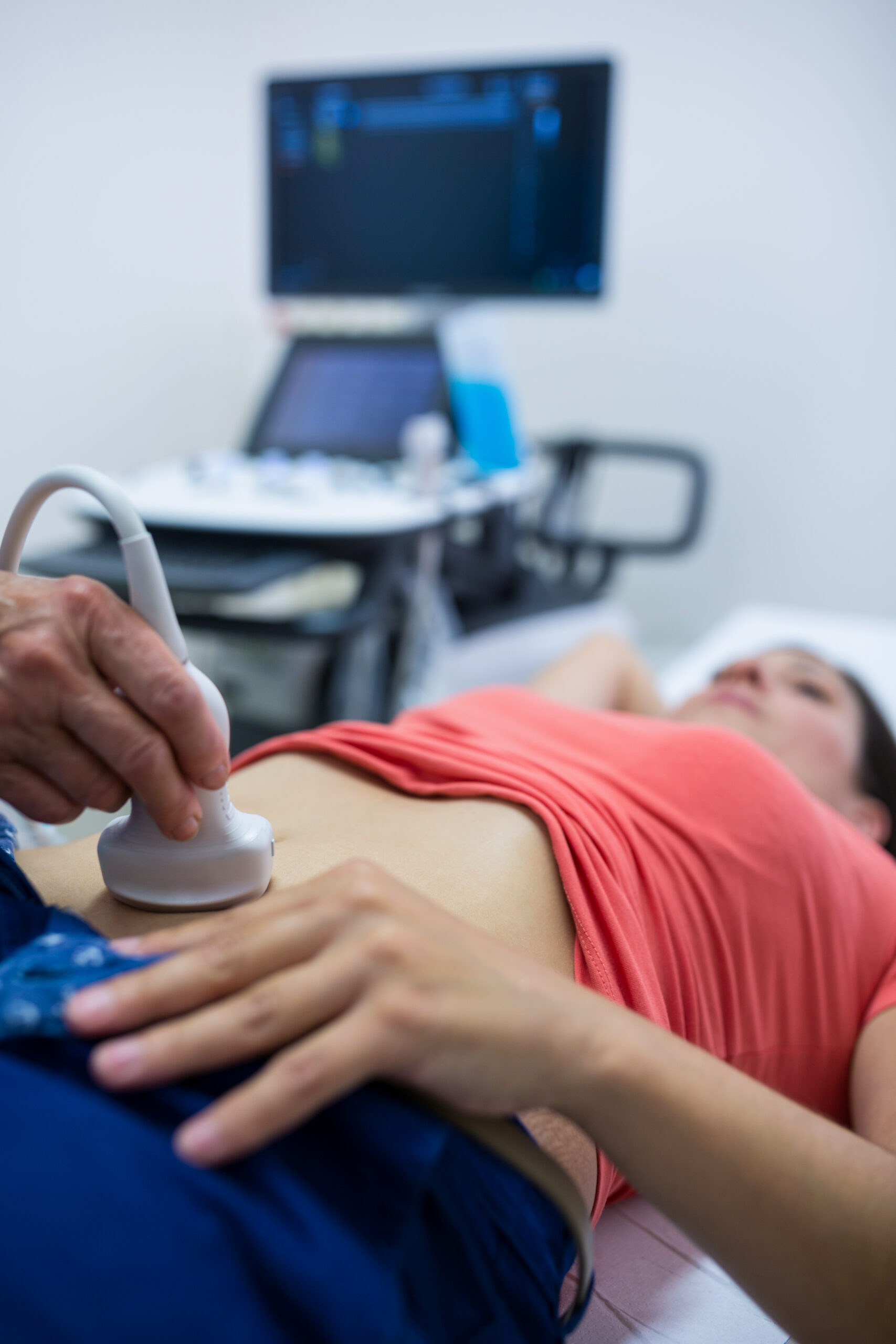
Breast Implant surgery is the top cosmetic surgery performed nowadays.It is a positive step for most women in which the number of women undergoing breast implant procedures is increasing rapidly. Yet, studies have shown that breast augmentation which is also known as breast implant can help boost self-esteem, body image,etc…
Moreover it can be done for reconstructive purposes such as after mastectomy for breast cancer, or for cosmetic reasons.
It is therefore imperative for the plastic surgeon to be familiar with the diagnostic imaging studies that are used to evaluate implant integrity, detect abnormalities of the implant and its surrounding capsule, and detect breast conditions unrelated to implants.
So which Ultrasound Scanner is the best for Breast Implant?
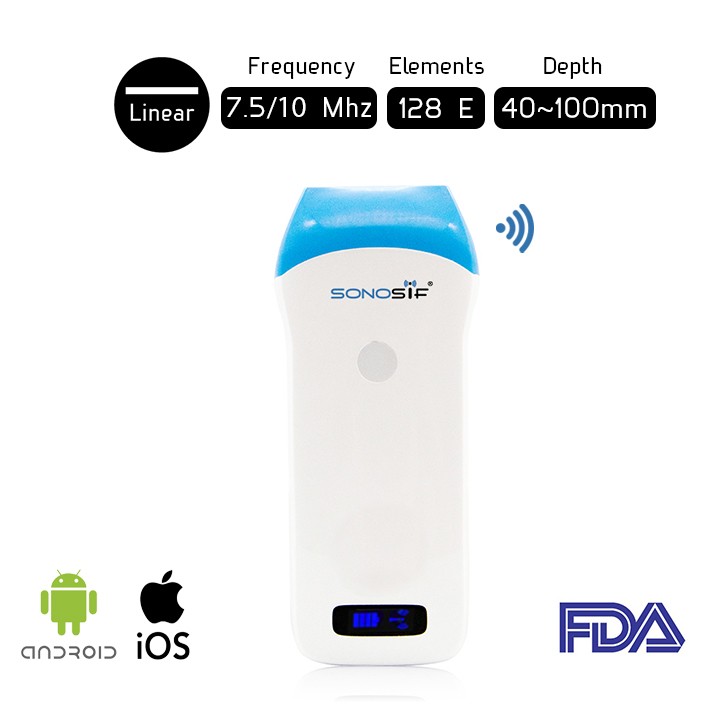
The use of Linear ultrasound scanner with high frequency from 7.5 to 10 MHz is vital for evaluating breast transplants including assessing the morphology, contour, content and peri-implant tissues and axillae.
More specifically, we recommend the Wireless Linear Ultrasound Scanner L7 – 7.5/10 Mhz to plastic surgeons and practitioners of aesthetic surgeries.
The Wireless Linear Ultrasound scanner L7-7.5/10 MHz provides an excellent visualization with its 256 level’s image gray-scale.
It helps the practitioner to surpass challenges such as the identification of the type of the implant, diagnosis of implant-related complications, as well as the diagnosis and follow-up of additional breast lesions such as cancer.
There are several risk factors that are related to the patient and the implant. In fact, up to 6% of cases show asymptomatic ruptures, most ruptures are contained without extension beyond the fibrous capsule.
The rupture of saline/silicone implants is associated with complete or immediate collapse of the cover and its surrounding fibrous capsule is easily recognizable by the doctor and is evident in the ultrasound and mammography.
In other words Liner ultrasound with depth from 40 mm to 100 mm is needed for post-surgical complications such as infection, implant leakage or rupture,The formation of tight scar tissue around the implant (capsular contracture),fluid accumulation (seroma), and hematoma,etc…
To sum up, ultrasonography and mammography are the first and last diagnostic tools that must be used in evaluating breast implants.
References : Breast implants, Asymptomatic ruptures, Risks of breast augmentation
Disclaimer: Although the information we provide is used by different doctors and medical staff to perform their procedures and clinical applications, the information contained in this article is for consideration only. SONOSIF is not responsible neither for the misuse of the device nor for the wrong or random generalizability of the device in all clinical applications or procedures mentioned in our articles. Users must have the proper training and skills to perform the procedure with each ultrasound scanner device.
The products mentioned in this article are only for sale to medical staff (doctors, nurses, certified practitioners, etc.) or to private users assisted by or under the supervision of a medical professional.


