Abdominal Aortic Aneurysm: AAA
September 19, 2020
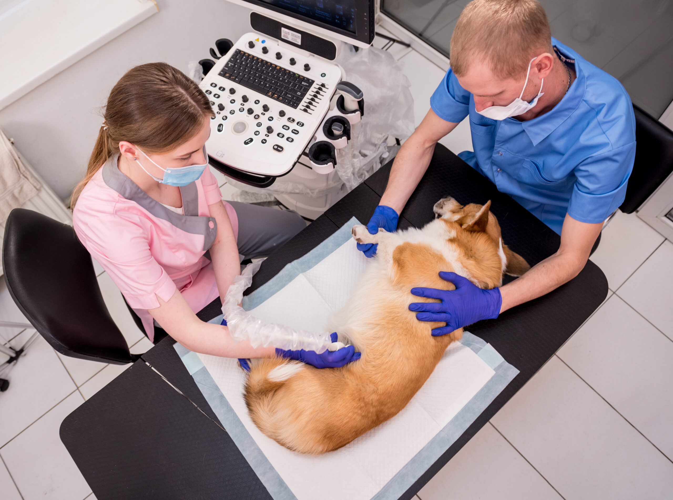
Pets Ultrasound Scanning
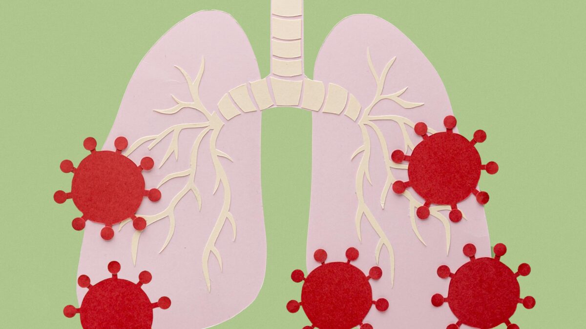
September 22, 2020COVID-19 is an infectious disease caused by the most recently discovered coronavirus. This new virus was first detected in Wuhan, China, in December 2019. COVID-19 is now a pandemic affecting many countries globally. It has infected nearly 30.000.000 people around the world.
People with COVID-19 have had a wide range of symptoms reported – ranging from mild symptoms to severe illness. Symptoms may appear 2-14 days after exposure to the virus. Such as fever or chills, cough, shortness of breath, fatigue, muscle or body aches, the new loss of taste or smell, sore throat, vomiting, congestion or runny nose, headache, and diarrhea. Yet, some people might be infected but only have very mild symptoms.
As lung abnormalities may develop experts have recommended early chest computerized tomography (CT) for screening suspected patients.
Moreover, Lung Ultrasound Scanner allows a bedside examination of patients, even those who are critically ill, without needing to transfer them. Thus, it could represent a valuable approach for the diagnosis and follow-up of lung involvement in COVID patients, minimizing the risk of further infection in healthcare personnel.
So which ultrasound scanner is the best for COVID-19 Lung Ultrasound?
Since lung ultrasound may aid in the identification and subsequent monitoring of suspected COVID-19 infections, perhaps even before the onset or progression of respiratory symptoms.
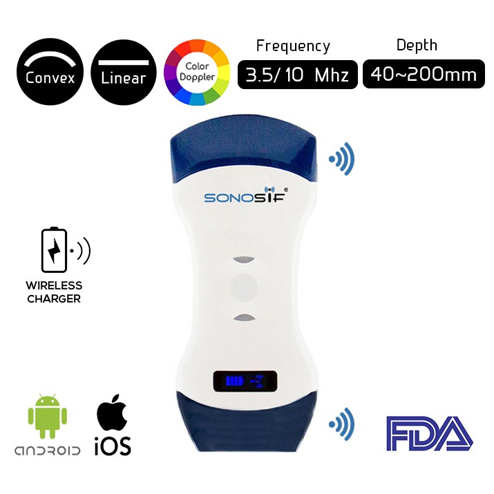
Our SONOSIF ‘s Research and Development team recommends the Convex and Linear Color Doppler WiFi Double Head Ultrasound Scanner CLCD to pulmonologists. Such a device minimizes the risk of contamination of devices and subsequent nosocomial spread.
The US CLCD is connected with a tablet and most importantly it is portable in which it is easy to sterilize due to smaller surface areas.
During the procedure, only two operators are needed, a doctor and a medical caretaker. In the isolation room respecting all the preventive measures for respiratory, droplet, and contact isolation provided by the World Health Organization for the new COVID-19 outbreak.
The ultrasound probe and the tablet were placed in two distinctive sterile, plastic probe, and tablet covers. The doctor performs lung ultrasound utilizing the wireless probe and, subsequently, entering in contact with the patient. The medical caretaker considers the tablet and is capable to freeze and store pictures/recordings, along these lines not contacting neither the patient nor whatever else inside the room.
Toward the end of the procedure, the tablet and test can be sanitized in a committed zone and put into two new clean plastic packs.
The advantages of ultrasonography, apart from the possibility of the bedside examination without the necessity of transporting the patient, include non-invasiveness and lack of exposure to X-rays.
Consequently, this examination can be performed as often as is clinically necessary. This may be particularly important for patients in very serious clinical conditions, who require advanced therapeutic techniques at Intensive Care Units (invasive mechanical ventilation, renal replacement therapy, extracorporeal membrane oxygenation – ECMO).
Transportation of such patients to tomography units to assess the development of pulmonary lesions and its pace may be risky or impossible. In this patient group, chest ultrasound, extended with basic echocardiography, assessment of the abdominal cavity and inferior vena cava (IVC) may be very useful in treatment monitoring and optimization.
To conclude, Lung Ultrasound Scanner is useful in the diagnosis of patients suspected of COVID-19 and it may be utilized for monitoring of the disease at Intensive Care Units (ICU).

References: COVID-19, Symptoms of COVID-19, Lung ultrasound in the diagnosis of COVID-19


