- Immediate contact :
- +1-323-988-5889
- info@sonosif.com

Pharyngitis Ultrasound Diagnosis
October 11, 2022
Monitoring IUD Insertion Ultrasound
October 31, 2022The release of an egg from its follicle in one of a woman’s two ovaries is one of the most important milestones in childbirth. When an egg is ovulated, it is picked up by one of the fallopian tubes and begins its trip toward the uterus.
Ovulation is triggered by an increase in luteinizing hormone (LH) levels in the woman’s blood and occurs 36 hours after the LH surge begins. When an egg is fertilized and implants in the endometrium, a pregnancy is formed. The endometrial lining that grows in preparation for pregnancy is shed as menstrual flow if a pregnancy does not occur.
Since a variety of factors can obstruct or disrupt ovulation and lead to infertility, evaluating whether or not a woman is ovulating is usually necessary.
Which Ultrasound Is the most suitable for ovulation detection?
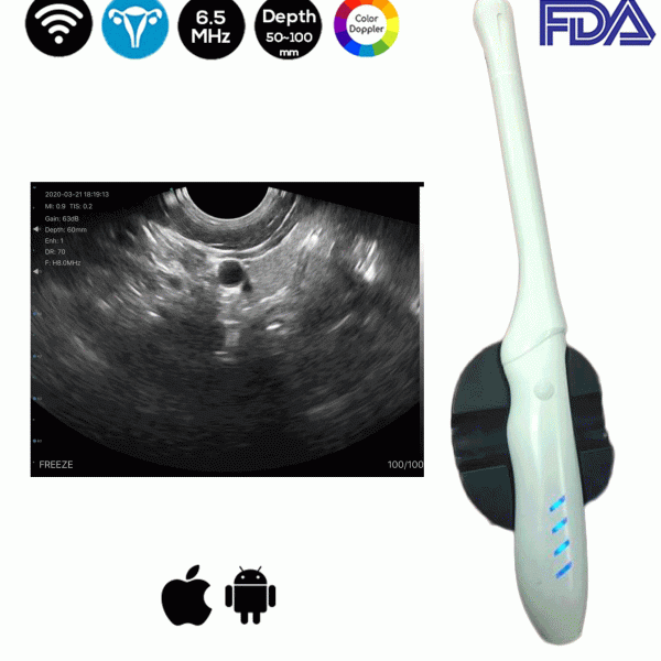
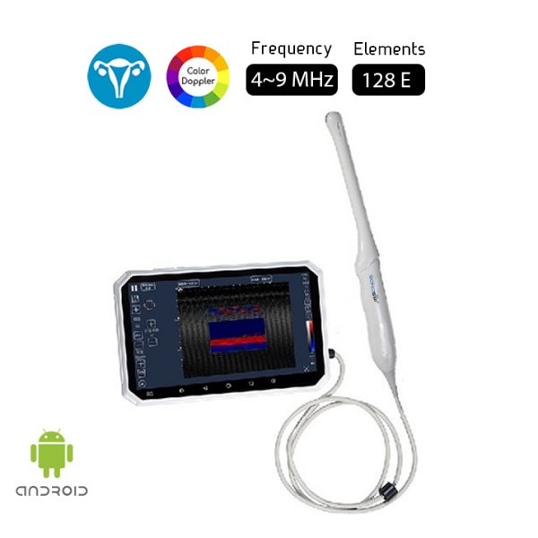
Ultrasonography imaging is a noninvasive, effective, simple, safe, and widely available method for evaluating and detecting ovulation. Our medical technical team recommends the Convex and Transvaginal Color Doppler Double Head Wireless Ultrasound Scanner CTC-3.1 for OB/GYN applications.
Ultrasound, a technology that employs sound waves to create a picture on a monitor screen, can be used to detect follicular development. This is a painless technique that is often performed with a probe put into the vagina, but it may also be performed with an external probe placed on the belly. The follicle is thin-walled and filled with fluid before ovulation. As the egg inside the follicle develops, so does the follicle. Ovulation usually occurs when the follicle is between 1.8 and 2.5 millimeters in diameter.
Ultrasound can aid in the timing of your intercourse or insemination. Ultrasound may be conducted on numerous distinct days during the menstrual cycle in women using fertility medications to correctly quantify and track each follicle.
Reference: Ultrasound scanning of ovaries to detect ovulation in women
Disclaimer: Although the information we provide is used by different doctors and medical staff to perform their procedures and clinical applications, the information contained in this article is for consideration only. SONOSIF is not responsible neither for the misuse of the device nor for the wrong or random generalizability of the device in all clinical applications or procedures mentioned in our articles. Users must have the proper training and skills to perform the procedure with each ultrasound scanner device.
The products mentioned in this article are only for sale to medical staff (doctors, nurses, certified practitioners, etc.) or to private users assisted by or under the supervision of a medical professional.


