Ultrasound-Guided Anserine bursa
May 6, 2021
Ultrasound-Guided Medial Collateral Ligament
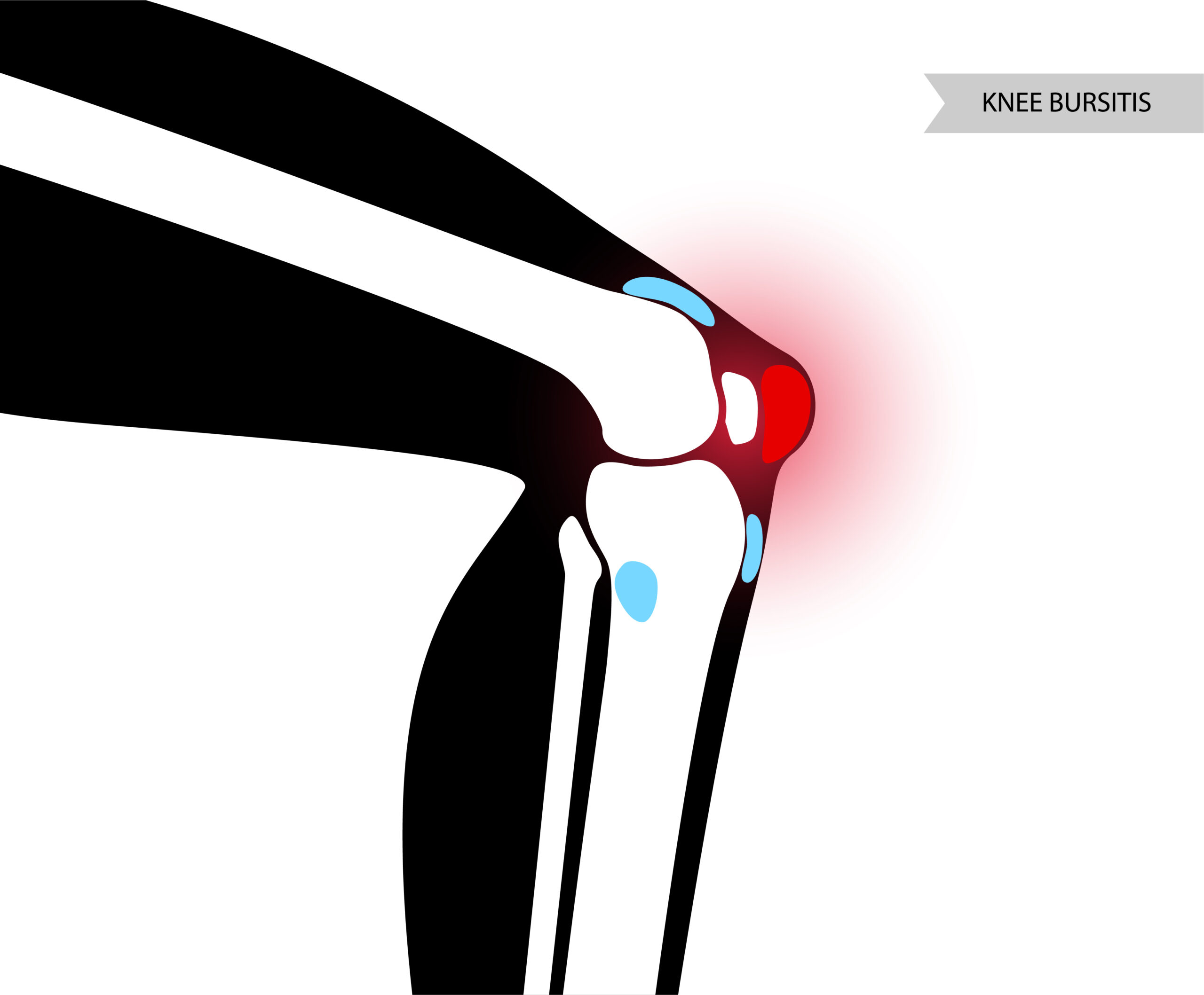
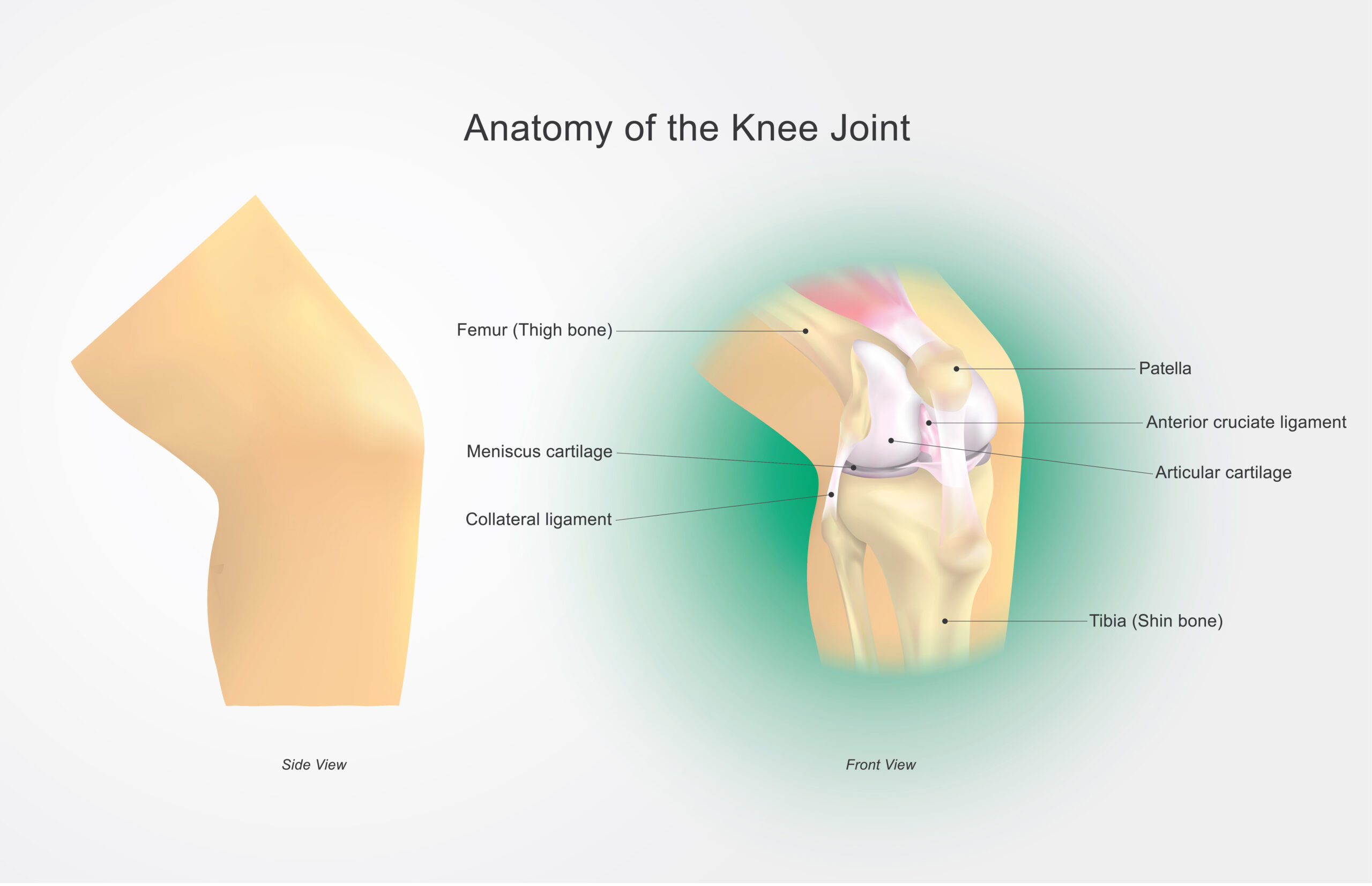
May 8, 2021The infrapatellar bursa lies deep between the patellar tendon and the upper front surface of the tibia or shin bone. Its function is to aid movement by lubricating the tendon as it moves over the bone.
It is made up of two sacs:
* Superficial Infrapatellar Bursa: lies between the anterior subcutaneous tissues of the knee and the anterior surface of the patellar tendon
* Deep Infrapatellar Bursa: lies between the patellar tendon and the shin bone (tibia), it is characterised by inflammation of the bursal synovium and associated with the formation of an increased amount of fluid and collagen.
Infrapatellar bursitis is characterized by discomfort and swelling in the front of the leg, just under the knee cap. It occurs when one of the thin fluid-filled sacs in the knee becomes irritated and inflamed.
Infrapatellar bursitis symptoms consist of pain at the front of the knee, Patients will likely have swelling over the area of the infrapatellar bursa. Its symptoms may be similar to that of jumper’s knee or patellar tendonitis with pain just below the kneecap.
Ultrasound imaging allows doctors to assess the Infrapatellar area of which it shows: deep, distended, cystallized underpateler bursa, internal septations and heterogeneous weight of soft tissue.
Which Ultrasound Scanner is the best in the assessment of Superficial and deep Infrapatellar bursa?
In the measurement of the infrapatellar bursa, orthopedists prefer to use a high-frequency linear transducer. For the improving point of treatment, clinical testing techniques and quality of clinical diagnose, the USB Linear 5-12MHz Ultrasound Scanner USB-UL2 plays a vital role.
It guides the diagnosis and help delineate the presence of another knee bursitis, calcific tendonitis, tendinopathy, patellar tendonitis, or other knee pathology.
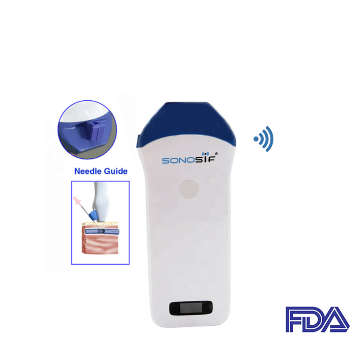
The Mini Linear Handheld WiFi Ultrasound Scanner MLCD can also be used by physicians to provide dynamic images of superficial soft tissues in real-time and also for ultrasound injections.
MLCD has a needle guide holder. As a result, it can be sent directly to the guidance pin frame. Combined with tools that can quickly determine the extent and diameter of a puncture. The latter enables the doctor to see the needle as it approaches the body and travels to the target position in real-time.
Ultrasonography is useful for detecting chronic fibrotic changes in the infrapatellar region, as well as synovitis-related inflammation. The use of Color Doppler US to visualize the inflammation inside the fat pad is effective.
References: Infrapatellar bursitis, Ultrasound-Guided Injection Technique for Deep Infrapatellar Bursitis Pain, Ultrasound-Guided Injection Technique for Superficial Infrapatellar Bursitis Pain, Deep infrapatellar bursitis,
Disclaimer: Although the information we provide is used by different doctors and medical staff to perform their procedures and clinical applications, the information contained in this article is for consideration only. SONOSIF is not responsible neither for the misuse of the device nor for the wrong or random generalizability of the device in all clinical applications or procedures mentioned in our articles. Users must have the proper training and skills to perform the procedure with each ultrasound scanner device.
The products mentioned in this article are only for sale to medical staff (doctors, nurses, certified practitioners, etc.) or to private users assisted by or under the supervision of a medical professional.


