- Immediate contact :
- +1-323-988-5889
- info@sonosif.com
Ultrasound-Guided Lateral and Medial epicondylitis
April 20, 2021
Ultrasound-Guided Suprapatellar recess
April 29, 2021The ankle joint allows up-and-down movement of the foot. The subtalar joint sits below the ankle joint and allows side-to-side motion of the foot. Numerous ligaments (made of tough, moveable tissue) surround the true ankle and subtalar joints, binding the bones of the leg to each other and to those of the foot.
The ankle is a large joint made up of three bones:
- The shin bone (tibia)
- The thinner bone running next to the shin bone (fibula)
- A foot bone that sits above the heel bone (talus)
Tendons traverse the anterior, posterior, and lateral aspects of the ankle. This involves the tibialis anterior, tibialis posterior, and a pair of peroneal tendons (peroneus longus and brevis). Because of their critical roles during gait, each of these tendons is prone to overuse and inflammation.
Ankle sprains, nerve injury, anterior process fracture in the calcane, or the base fracture of the fifth metatarsal, can cause lateral ankle pain. Though navicular accessories, spring ligament injuries, or medial malleolar stress fractures can all cause medial ankle pain close to the back of the tibialis.
While various imaging modalities can be used to diagnose ankle pain, ultrasound (US) has several advantages for evaluating ankle pain, particularly in the tendons, ligaments, and nerves of the ankle.
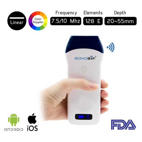
A high-frequency linear transducer with a frequency range of 7.5 to 10 MHz is appropriate for assessing tendons in the medial and lateral compartments of the ankle. For this reason, our medical research and development team always recommends the Wireless Color Doppler Linear Ultrasound Scanner L2CD to our orthopedist clients.
TheL2CD is a compact handheld scanner with advanced features. This improves the physical examination and gives the physician great help during the evaluation. It has the colour Doppler mode which helps to distinguish small intra-substance tears from blood vessels that can occur in a tendinopathic tendon.
During the Tendons assessment and follow-up stage, the Ultrasound Scanner is highly valuable. It advances knowledge and research into traumatic distortion of the ankle’s capsule-ligament structures, as well as degenerative, infectious, and traumatic lesions of the ankle tendons.
References: The ankle , Tendon Disorders of the Foot and Ankle, Ultrasonography of the ankle joint,Medial ankle ligament,
Disclaimer: Although the information we provide is used by different doctors and medical staff to perform their procedures and clinical applications, the information contained in this article is for consideration only. SONOSIF is not responsible neither for the misuse of the device nor for the wrong or random generalizability of the device in all clinical applications or procedures mentioned in our articles. Users must have the proper training and skills to perform the procedure with each ultrasound scanner device.
The products mentioned in this article are only for sale to medical staff (doctors, nurses, certified practitioners, etc.) or to private users assisted by or under the supervision of a medical professional.


