- Immediate contact :
- +1-323-988-5889
- info@sonosif.com

Ultrasound Scanning for Paget’s Disease of the Bone
April 21, 2022
Ultrasound-Guided Phlebotherapy (TRAP)
April 23, 2022Paget’s disease of the nipple, also known as Paget’s disease of the breast, is a rare condition associated with breast cancer. It causes eczema-like changes to the skin of the nipple and the area of darker skin surrounding the nipple (areola). It’s usually a sign of breast cancer in the tissue behind the nipple.
Doctors don’t know what causes Paget’s disease of the breast. The most widely accepted theory is that the disease results from underlying ductal breast cancer. The cancer cells from the original tumor then travel through milk ducts to the nipple and its surrounding skin.
The following signs are the most common symptoms of Paget’s nipple disease:
· Itching, tingling, or redness in the nipple and/or areola.
· Flaking, crusty, or thickened skin on or around the nipple.
· A flattened nipple.
· Discharge from the nipple that may be yellowish or bloody.
One of the possible and most practical ways of diagnosis is ultrasonography. Ultrasound is used not only to confirm the mammography findings in the case of Paget’s disease but also when the mammogram is negative. The findings on ultrasound include heterogeneous hypoechoic areas in breast parenchyma and the presence of a discrete mass or dilated ducts.
To be really able to detect all these issues in a highly professional way providing top-quality scan imagery, a qualified scan machine should be employed.
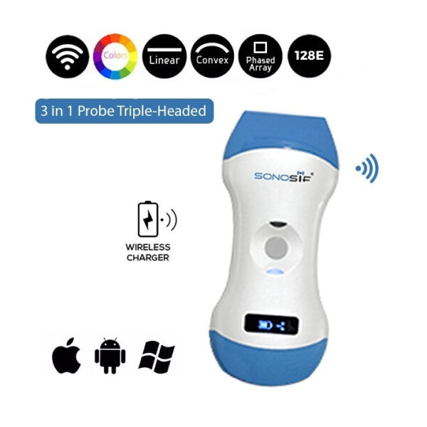
In this regard, the Color Doppler 3 in 1 Wireless Ultrasound Scanner 3in1-CLC1CD, a top recommendation from mammography specialists, met the ultrasonic criteria required for this specific scanning process.
The following wireless ultrasound scanner provides qualitative and quantitative mammographic data thanks to its smaller footprint it aids in the evaluation of the breasts’ soft tissues and even nerves. Further, this multi-use ultrasound scanner is also a highly effective POC ultrasound scanner. That is, It can easily and safely be used by ordinary individuals suspecting any-breast related issue before and after a doctor’s actual exam to maintain free gradual check-ups.
Speaking more about this handheld ultrasound machine’s specificities, it functions through 3 major Working Systems: Apple iOS and Android, and Windows which means it is compatible with both your PC and smartphone to ensure complete diagnosis transparency. The mobile ultrasound scanner is also rechargeable by a Wireless Charger.
Still, the most exquisite feature of this portable ultrasound is yet to come. It is able to provide puncture assistance with an accurate Needle Point Guide which might be greatly needed during operations.
In sum, the 3in1-CLC1CD ultrasound machine is an excellent wireless ultrasound scanner for mammographic medical establishments in particular. The reason is that It does not require any extra training to use due to its simple interface. It’s light, portable, and simple to operate. But, most importantly, it is ideal for Paget’s nipple patients who have started to notice certain breast cancer signs and are still not sure of its existence or at which stage the disease is. These individuals surely need a very precise diagnosis leading to successful treatment as fast as it could be.
Reference: Paget’s disease of the nipple
Disclaimer: Although the information we provide is used by different doctors and medical staff to perform their procedures and clinical applications, the information contained in this article is for consideration only. SONOSIF is not responsible neither for the misuse of the device nor for the wrong or random generalizability of the device in all clinical applications or procedures mentioned in our articles. Users must have the proper training and skills to perform the procedure with each ultrasound scanner device.
The products mentioned in this article are only for sale to medical staff (doctors, nurses, certified practitioners, etc.) or to private users assisted by or under the supervision of a medical professional.


