- Immediate contact :
- +1-323-988-5889
- info@sonosif.com

Ultrasound-guided Caudal Injection
May 26, 2021
Infertility Ultrasound
June 1, 2021Ejection fraction (EF) is a measurement of how much blood the left ventricle pumps out with each contraction, represented as a percentage. With each heartbeat, 60% of the blood in the left ventricle is pushed out.
Which Ultrasound Scanner is the best for Ejection Fraction measurement?
Ejection Fraction is usually assessed in the left ventricle because it does the most of the work in the body. However, recent study suggests that while evaluating EF, the right ventricle should not be overlooked.
A number of imaging modalities can be used to provide a precise reading of the left ventricular Ejection Fraction.
The echo-cardiogram, which employs sound waves to produce pictures of the heart, is one of the most frequent EF testing methods.
To illustrate how blood flows between the heart valves, echo examinations are frequently paired with Doppler imaging equipment.
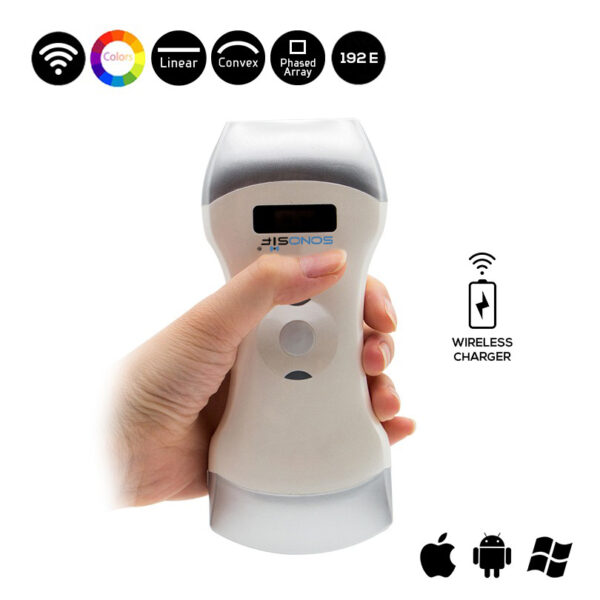
Our medical Research and Development team constantly recommends the 3 in 1 Color Doppler Wireless Ultrasound Scanner 3in1-CLC3CD to cardiologists and primary care doctors.
The 3in1-CLC3CD produces clear pictures of the heart’s chambers and valves using high-frequency sound waves ranging from 3.5 MHz to 7.5 MHz, allowing clinicians to observe how the heart is working.
Indeed, studies have shown that Research shows that the volume of blood within a ventricle at the end of the diastole is the end-diastolic volume (EDV). Similarly, the end-systolic volume is the amount of blood remaining in a ventricle at the end of systole (contraction) (ESV). The stroke volume is the difference between EDV and ESV (SV).
The ejection fraction is inherently a relative measurement—as is any fraction, ratio, or percentage, whereas the stroke volume, end-diastolic volume, or end-systolic volume are absolute measurements.
The calculation of LVEF is important for diagnostic, prognostic, and therapeutic purposes, and a quick, accurate, repeatable, and non-invasive technique would be ideal.
Ultrasonography is highly needed during the measurement of ejection fraction, especially that Left ventricular ejection fraction (EF) is an important and sensitive parameter in the diagnosis and treatment of patients with coronary heart disease.
References: Ejection Fraction, The volume of blood, Ejection Fraction measurement , Ejection Fraction Heart Failure Measurement,
Disclaimer: Although the information we provide is used by different doctors and medical staff to perform their procedures and clinical applications, the information contained in this article is for consideration only. SONOSIF is not responsible neither for the misuse of the device nor for the wrong or random generalizability of the device in all clinical applications or procedures mentioned in our articles. Users must have the proper training and skills to perform the procedure with each ultrasound scanner device.
The products mentioned in this article are only for sale to medical staff (doctors, nurses, certified practitioners, etc.) or to private users assisted by or under the supervision of a medical professional.


