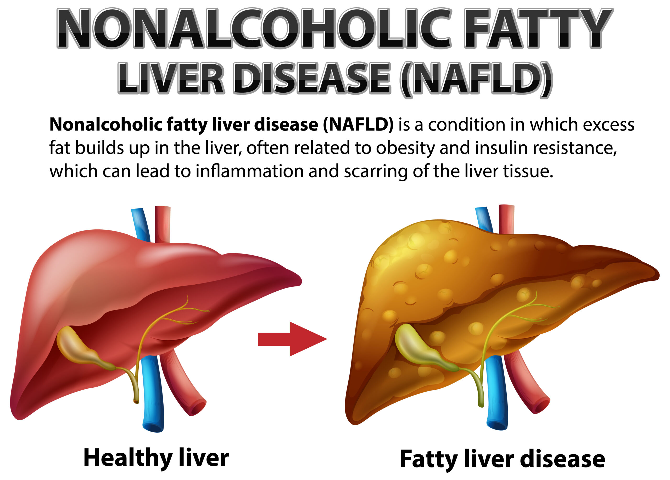
Nonalcoholic Fatty liver Disease Bedside Ultrasound Diagnosis
February 14, 2022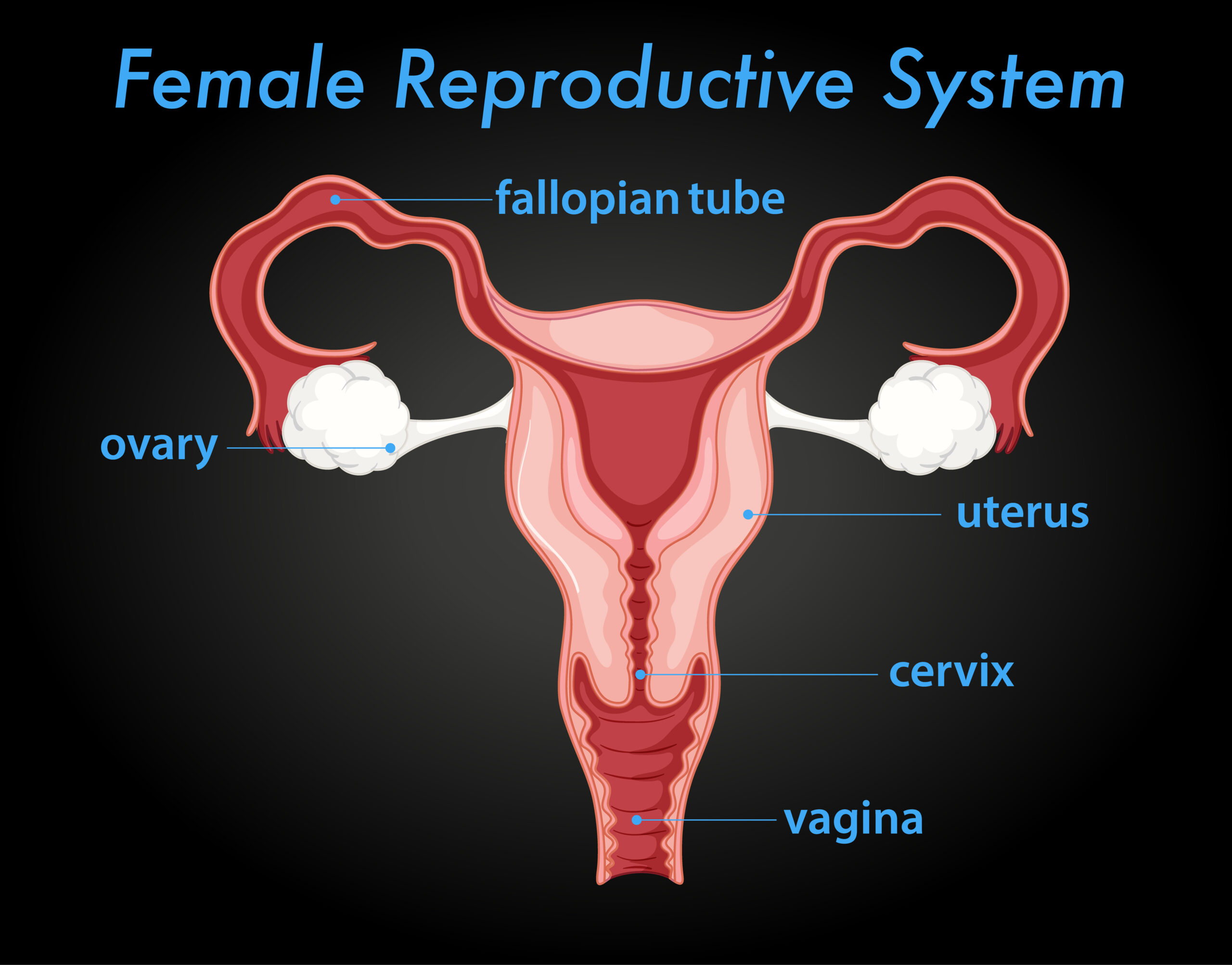
US Characterization and Reporting of Adnexal Masses
February 14, 2022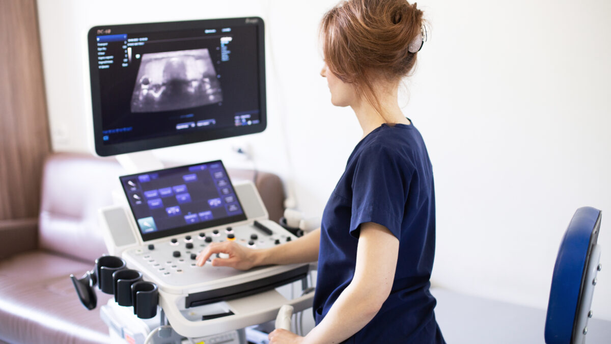
The World Health Organization defines telemedicine as “the delivery of health care services where distance is a critical factor.”
Telemedicine services are intended for the exchange of accurate information for disease diagnosis, prevention, and treatment, as well as continuing education for health care providers, as well as research and evaluation.
Ultrasonography is a valuable diagnostic tool because it is non-invasive, generally inexpensive, and highly portable, and it does not use ionizing radiation.
However, the generation and interpretation of ultrasound images are highly dependent on the operator. As a result, the performance and interpretation of these exams have traditionally been restricted to medical specialists.
Opinions in the literature are divided with regard to the transmission system involved in teleultrasonography. Some authors consider that good image quality can only be obtained with the use of asynchronous transmission, which, in addition to good diagnostic accuracy, allows for the training and professional supervision to produce a satisfactory level of clinical competence.
Other studies have sought to demonstrate the accuracy of the teleultrasonography performed in real-time between a tertiary center and a remote area. The authors argue that the image quality was not very clear when teleultrasonography first started but that current telecommunication and image compression technologies have made high-quality synchronous and asynchronous transmissions feasible.
Other authors argue in favor of real-time transmissions because asynchronous mode only allows images and videos to be stored for future analysis, and their interpretation may be incomplete or diagnostically inaccurate if some critical information is missing and cannot be recovered.
The authors also thought that this educational tool was superior to verbal instruction when training doctors at a distance because it allows new skills to be learned in half the time that traditional educational practices require.
This technology may be useful when a cardiologist is at home and a hospital is attempting to obtain a clear image of a patient who may or may not be suffering from heart failure. The cardiologist can log in, saving a trip to the hospital and preventing this critical exam from being missed.
Another example of the usefulness of this wonderful technology is in an emergency or trauma care situation where immediate assessment is required.
It is critical to be able to use ultrasound on a patient for a variety of applications (e.g., to identify the impact of trauma, the source and extent of the injury and the impact of treatment rendered) and then put together a plan for whether the patient needs to be transferred to a higher level of care or can remain in their home community. It can then be used as an educational tool for both the remote provider and future patients/cases.
The PW USB-L3CD Handheld Color Linear 128E Ultrasound Scanner, 5-10MHz is a new handheld medical imaging device that can significantly reduce the cost and efficiency of medical ultrasounds. With a special ultrasound scanner/transducers attached to a person’s chest, neck, abdomen, or any part of the body’s skin, this compact and versatile device the size of an iPhone 6 plus could be held to a person’s chest, neck, abdomen, or any part of the body’s skin. As a result, you can generate vivid, moving, and clear images of what’s inside in real-time.
The handheld Color linear 128E Ultrasound Scanner 5-10MHz PW USB-L3CD can upload images to the private cloud in real-time using a high-speed WiFi/Bluetooth data connection, integrating telemedicine into service, so that a specialist in a remote location can weigh in on the images that the device records.
The device will extract key features/characteristics in the image by combing through the bank of images. Wavelet filter, pseudocolor, image smoothing, frame correlation, 128E, and 5M pixel camera are all included in the PW USB-L3CD . Deep-learning artificial intelligence will enable this pocket-sized ultrasound to perform automated diagnoses in the future.
Reference: The Feasibility of Real-Time Transmission of Sonographic Images from a Remote Location over Low-Bandwidth Internet Links: A Pilot Study
The evidence base of telemedicine: overview of the supplement
-
Product on sale
 Color Doppler USB Linear Ultrasound Scanner 5-10Mhz PW USB-L3CDOriginal price was: $3,250.$2,685Current price is: $2,685.


