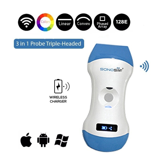
Color Doppler 3 in 1 Wireless Ultrasound Scanner 3in1-CLC1CD
November 25, 2019
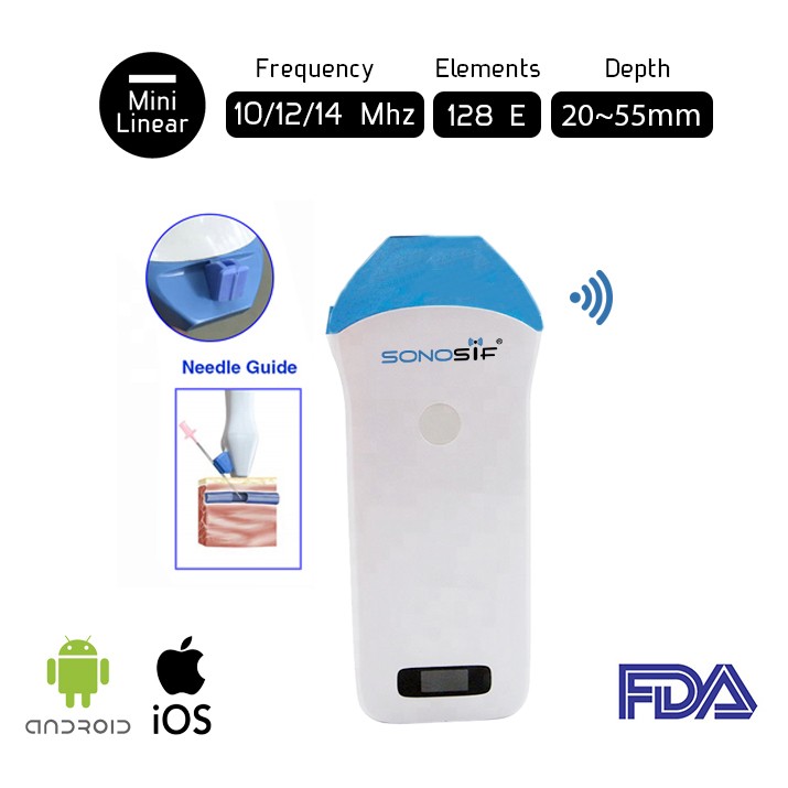
Built-in Screen Linear Wireless Ultrasound Scanner BiS-L1CD
November 25, 2019

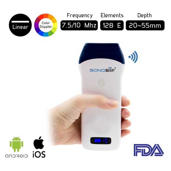
Color Doppler Wireless Linear Ultrasound Scanner L7CD
Original price was: $4,500.$3,598Current price is: $3,598.
Superior Image Quality
Wireless Freedom
Works on Android and iOS
Approvals : CE, ISO
Free Express Shipping |
Worldwide |
|---|---|
Warranty |
15 Months |
Return Policy |
7 Days |
Color Doppler Wireless Linear Ultrasound Scanner L7CD
An ultrasound scanner is the third eye of a doctor. The Color Linear Ultrasound Scanner L7CD is a wireless ultrasound scanner. It allows the doctor to make clinical applications anywhere, anytime. The L7CD has 5-10 MHz frequency and 40-120mm.
Also, the Color Linear Ultrasound Scanner’s image mode is B, M and color. The B-mode or brightness mode: produced by scanning the transducer beam in a plane as shown. It can be used for both stationary and moving structures such as cardiac valve motion. On the other hand, the M-mode or motion mode: it displays the A-mode signal corresponding to repeated pulses in a separate column of a 2-D image. It is mostly employed in conjunction with ECG for motion of the heart valves.

Consequently, the L7CD is the leading modality in vascular access. For instance, Vascular cannulation: Intravenous (IV) cannulation is a technique in which a cannula is placed inside a vein to provide venous access. Venous access allows sampling of blood, as well as administration of fluids, medications, parenteral nutrition, chemotherapy, and blood products. Further, this probe is used for calculation of the speed of blood flow in the vessel, valuation of blood flow in the arteries and veins of the body and to diagnose blood clots in the veins of the arms and legs.
The major advantage of ultrasound is the real-time availability that is particularly important for intraoperative imaging. Ultrasound is used as navigation tool in traditional surgical interventions and in guided biopsies, such as biopsy of breast tumors. The Color Linear Ultrasound Scanner provides great internal detail when assessing soft tissue structures such as tendons and nerves. It can show the movement of a soft tissue structure such as a tendon, joint or an extremity.
Besides, the Wireless Ultrasound Probe does not only serve cardiology but also orthopedy. The Color Linear ultrasound scanner provides qualitative and quantitative for musculoskeletal diagnosis. For example: Tendon tears, or tendinitis of the rotator cuff in the shoulder, Achilles tendon in the ankle and other tendons throughout the body, muscle tears, masses or fluid collections., ligament sprains or tears.
Using the LWC1 the physician can detect; inflammation or fluid (effusions) within the bursae and joints, early changes of rheumatoid arthritis, nerve entrapments such as carpal tunnel syndrome, benign and malignant soft tissue tumors, ganglion cysts, hernias., foreign bodies in the soft tissues (such as splinters or glass), dislocations of the hip in infants, fluid in a painful hip joint in children, neck muscle abnormalities in infants with torticollis (neck twisting), soft tissue masses (lumps/bumps) in children.
The Color Linear Ultrasound Scanner also is:
- Useful in reconstructive surgery for identification of perforators for a variety of flaps, (including the anterolateral thigh flap, the deep inferior epigastric perforator flap).

- Reduce tissue trauma and improve skin contraction.
- Panning lymphaticovenular anastomoses.
- Study the integrity and rotation of breast implants.
- The management of Breast Implant-Associated Anaplastic Large-Cell Lymphoma.
- The evaluation of breast masses, including those that occur after autologous fat grafting.
- To quantitate changes in fat volume after fat injection of the breasts and buttocks.
- To measure decreases in thickness after nonsurgical fat reduction including cryolipolysis.
- Evaluation of facial hyaluronic acid injection and subcutaneous thickness after botulinum toxin injection.
- Screen patients for abdominal wall defects before liposuction or abdominoplasty.
- To evaluate repairs of the rectus abdominis diastasis, and for seroma management.
- Visualizing tendons and foreign bodies of the upper extremities and guiding injections.
- Assisting surgeons who perform thoracic wall, paravertebral, and transversus abdominis plane nerve blocks.
Features:
- Linear probe.
- Frequency(MHz): 10MHz.
- Bandwidth(MHz): 5-14MHz.
- Length: 38mm.
- Image Adjust: Gain, Focus, Harmonic, Denoise.
- Application: Peripheral blood vessels, small part.
Specifications:
- Scanning method: Linear.
- Connection mode: WiFi with iOS and Android.
- Software support system: Android.
- Image mode: B,M,Color.
- Probe type: Linear array.
- Probe length: B,B/M,C,B+B.
- Frequency bandwidth: 5-10MHz.
- Elements: 128.
- Channel: 32.
- Power: built-in lithium battery.
- Max depth: 12cm.
- Measurement: distance, perimeter, area and advanced private measurement package.
- Parameter: depth, gain and denoise.
- Connection interface: micro USB.
- Storage function: pictures and videos.
- Duration of battery: B mode: 6Hrs.
- Weight: 290g.
- Others: using plane wave algorithm, even image.
Scan Result

Carotid Artery and Internal Jugular Vein Imaging
| Head Type | Linear. |
|---|---|
| Screen Mode | B,M,Color. |
| Frequency | 5 – 10MHz. |
| Depth | 12cm. |
| Elements | 128. |
| Applications | Plastic Surgery, Gynecology, Abdominall Wall, Subcutaneous Injection. |
Related products
-
Wireless Convex and Linear Double Head Ultrasound Scanner CL
Original price was: $3,000.$2,525Current price is: $2,525. -
Wireless Color Doppler Linear Ultrasound Scanner L2CD
Original price was: $3,200.$1,999Current price is: $1,999. -
Mini Linear Handheld Wireless Ultrasound Scanner ML1
Original price was: $3,500.$1,795Current price is: $1,795.


Reviews
There are no reviews yet.