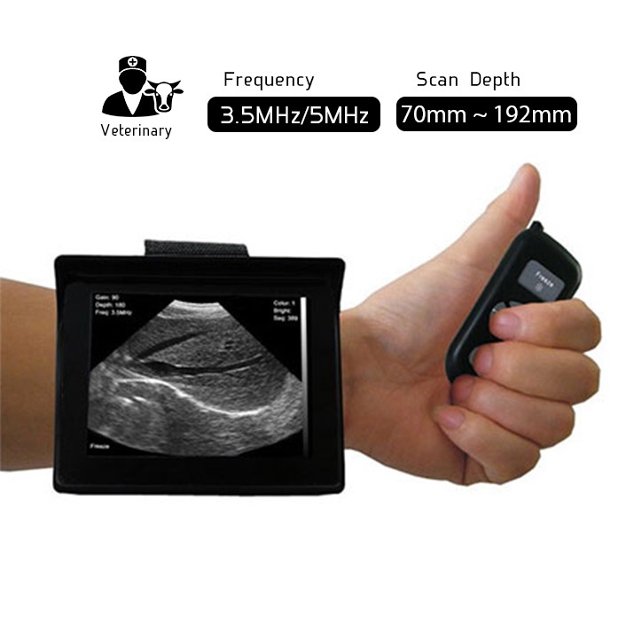
Portable Wrist-Wear Veterinary Ultrasound Scanner Vet-1
June 4, 2019
Wireless 3 in 1 Ultrasound Scanner Triple Headed: Convex, Linear and Cardiac Probe 3in1-CLC2CD
November 22, 2019
PW Doppler Super Width Linear Ultrasound Scanner SWL1CD
Original price was: $5,000.$2,895Current price is: $2,895.
Superior Image Quality
Wireless Freedom
Works on iOS and Android
Approvals : FDA, CE, ISO
Free Express Shipping |
Worldwide |
|---|---|
Warranty |
15 Months |
Return Policy |
7 Days |

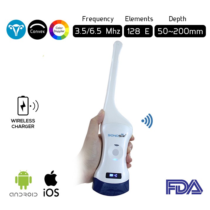
Super Width Linear Ultrasound SWL1CD has 256 elements. Thus, provides excellent resolution. Further, The probe has a 7.5 – 10 MHz offering visual depth up to 100mm. Thereof, the super width ultrasound scanner SWL1CD is suitable for breast and hip bone inspections.
For example, a plastic surgeon can use the super width ultrasound scanner, not only in reconstructive surgery for identification of perforators for a variety of flaps. – including the anterolateral thigh flap, the deep inferior epigastric perforator flap-. But also, in planning lymphaticovenular anastomoses.
In addition, the probe allows the practitioner to study the integrity and rotation of breast implants. Not to mention the management of Breast Implant-Associated Anaplastic Large-Cell Lymphoma. Besides that, the SWL1CD can come in handy in the evaluation of breast masses, including those that occur after autologous fat grafting.
Furthermore, the device offers qualitative and quantitative data. To allow the physician to quantitate changes in fat volume. After fat injection of the breasts and buttocks. Also, to measure decreases in thickness after nonsurgical fat reduction including cryolipolysis.

Moreover, the SWL1CD has proven its efficiency in the evaluation of facial hyaluronic acid injection and subcutaneous thickness after botulinum toxin injection. And its ability in visualizing tendons and foreign bodies of the upper extremities and guiding injections.
Thanks to its portability and light weight, the device is great for day-to-day procedures. Such as, screening for abdominal wall defects before liposuction or abdominoplasty. To evaluate repairs of the rectus abdominis diastasis, and for seroma management, assisting surgeons who perform thoracic wall, paravertebral, and transversus abdominis plane nerve blocks.

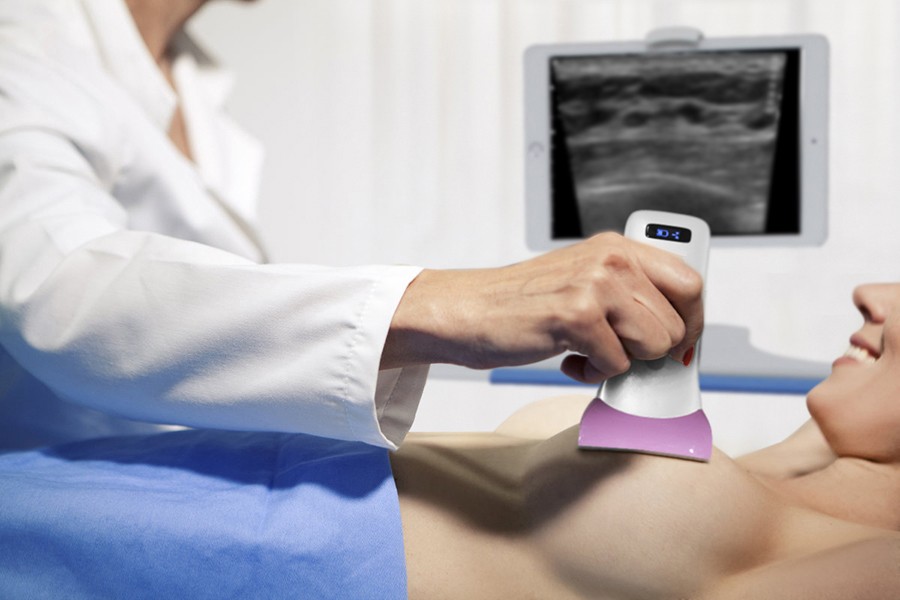
Breast exams
Super Width Ultrasound Probe in Breast exams:
On the other hand, the super width ultrasound scanner provides pictures of muscles, tendons, ligaments, joints, bone and soft tissues of the hip. In infants, the hip (which has a ball and cup configuration) is composed mainly of cartilage and is easily recognized on ultrasound.
 Hip ultrasound images using the linear probe are typically used to help evaluate:
- Abnormalities of the muscles, such as tears and soft-tissue masses.
- Foreign bodies, bleeding, infections or other types of fluid collections.
- Benign and malignant soft tissue tumors.
- Early changes of arthritis.
- Infant ultrasound can be used to check the hips for developmental dysplasia of the hip (DDH), which in infants can range from a shallow cup (bony acetabular dysplasia), to complete dislocation with the ball of the femoral head completely outside the socket.
Nonetheless, a doctor may as well use the super width ultrasound scanner if a suspicious lump is discovered in your breast. The ultrasound helps doctor determine whether the lump is a fluid-filled cyst or a solid tumor. It also allows them to determine the location and size of the lump.
Features :
- Works on iOS or Android (Tablet or Smartphone).
- Built-in and replaceable battery.
- Advanced digital imaging technology, superior image quality (256 Elements).
- High cost-effective.
- Wireless connectivity, easy to operate.
- Small light and easy to carry.
- Intelligent terminal platform, powerful expansion functions on application, storage, communication and printing.
Specifications :
- Item: Super Width Ultrasound Scanner.
- Scanning mode: electronic array.
- Display mode: B, B/M, B+Color, B+PDI, B+PW.
- Channel of RF circuit board: 16/32/64.
- Frequency: 7.5MHz/10MHz.
- Probe element: 256.
- Head width: 80mm.
- Scan depth: 20/40/60/100mm.
- Image Adjust: BGain, TGC, DYN, Focus, Depth, Harmonic, Denoise, Color Gain, Steer, PRF.
- Cineplay: auto and manual, frames can set as 100/200/500/1000.
- Puncture assist function: the function of in-plane puncture guide line, out-of-plane puncture guide line, automatic blood vessel measurement.
- Measure: Length, Area, Angle, heart rate, Obstetrics…
- Image save: jpg, avi and DICOM format.
- Image frame rate: 18 frames / second.
- Battery working time: 3~5 hours(according to different probe and whether keep scan).
- Battery charge: by USB charge or wireless charge, take 2 hours.
- Dimension: 156×60×20mm.
- Weight: 220g.
- Wifi type: 802.11g/20MHz/5G/450Mbps.
- Working system: Apple iOS and Android, Windows.
Scan Result:


| Head Type | Linear probe |
|---|---|
| Scan mode | Electronic array |
| Frequency | 5 MHz – 10 MHz |
| Image frame rate | 12f/s. |
| Depth | 0-100mm |
| Applications | Thyroid, carotid, breast, MSK, Vascular, Superficial, lung |
Related products
-
Wireless 4D Bladder Ultrasound Scanner B-4D FDA
Original price was: $3,000.$2,095Current price is: $2,095. -
Convex and Linear Color Doppler wireless Double Head Ultrasound Scanner CLCD
Original price was: $5,000.$2,895Current price is: $2,895. -
Wireless Linear Mobile Ultrasound Scanner L7 – 7.5/10 Mhz
Original price was: $2,350.$1,649Current price is: $1,649.


Reviews
There are no reviews yet.